Park City, UT

3 days / 6 sessions
Current Issues in Spine

February 2-4, 2017

Screen-Shot-2017-01-19-at-11.35.09-PM-1200x673.png

January 20, 2017 OrthoSpineNews


PROXIMAL HUMERUS 3-DIMENSIONAL FRACTURE MANAGEMENT SYSTEM

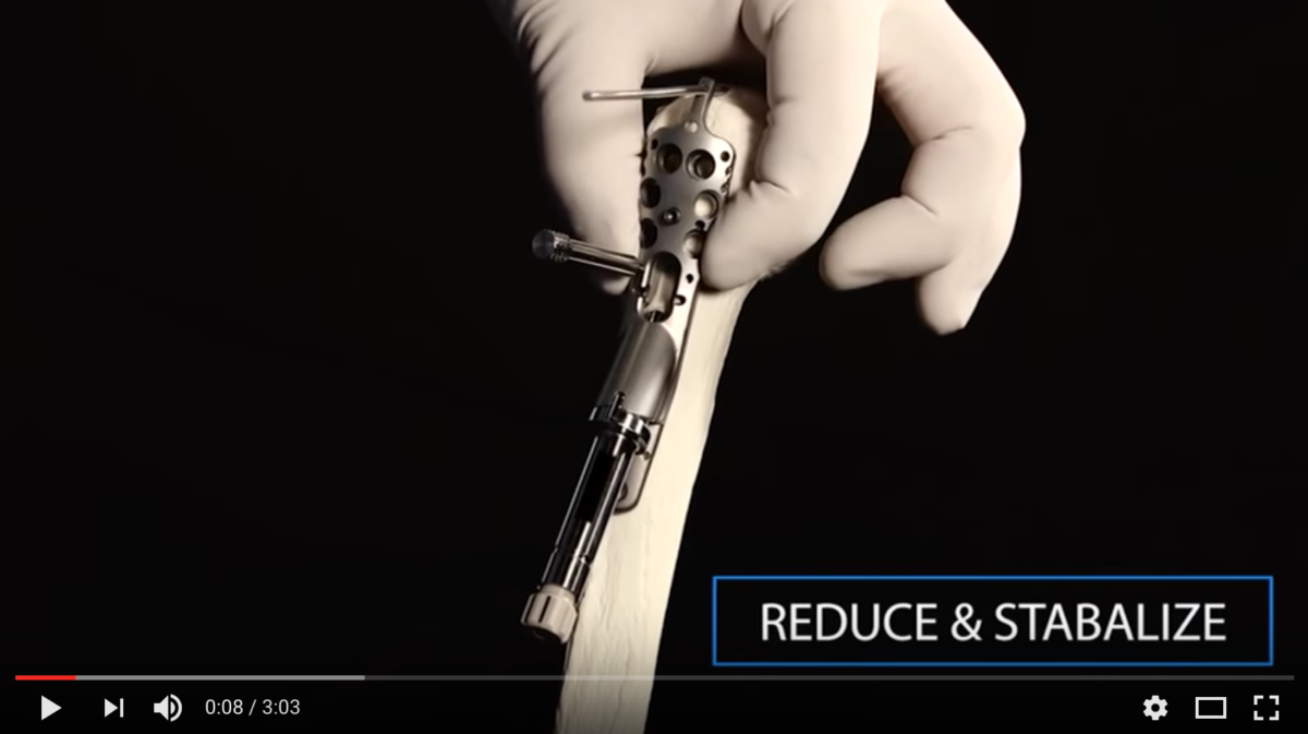
Proximal humerus fractures have remained a challenge to the orthopedist. No clear repair technology or approach has proven to be preferred in clinical literature. Correct anatomic reduction, medial column support, and bone mineral density are the leading contributors to failures for these patients. The Conventus PH Cage™ is the industry’s first alternative designed to directly address these modes of failure with a new technology that provides intramedullary buttress for fixation.

MEDIAL COLUMN SUPPORT

The Cage provides direct medial column support by placing a specifically engineered structural component in the intramedullary space and anchoring this device to the diaphyseal column of the humerus. This approach is a similar idea to using fibular struts and other materials to address medial column support in humerus fractures. The Cage provides a device that is properly shaped and sized for each patient, and removable should the need arise.
Screen-Shot-2017-01-19-at-11.29.58-PM-1200x667.png

January 20, 2017 OrthoSpineNews

EIT Cellular Titanium®

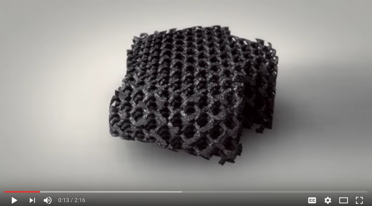
  • EIT Cellular Titanium® implants are produced with Selective Laser Melting (SLM) technique
  • EIT Cellular Titanium® consists of  ~ 80% porosity and a diamond pore size of ~ 650 μm, mimicking trabecular bone structure
    » Bone grafting is not necessary
  • Combination of solid and cellular implant architecture facilitates the rebuilding of natural cortical and cancellous bone structure
    » Provides optimal biomechanical and biological environment for natural bone ingrowth
  • Hydrophilic EIT Cellular Titanium®
    » Maximized blood contact leads to accelerated protein and mesenchymal cell attachment and bone cell differentiation
    » Proven biocompatibility of titanium alloy TiAl6V4
  • Excellent imaging characteristics
    » Implant contours visible under x-ray. Fusion area clearly visible due to high implant porosity
    » MRI and CT compatible


Screen-Shot-2017-01-19-at-11.21.32-PM-1200x676.png

January 20, 2017 OrthoSpineNews


UNiD Process

Unid-process-01

1.PLAN   
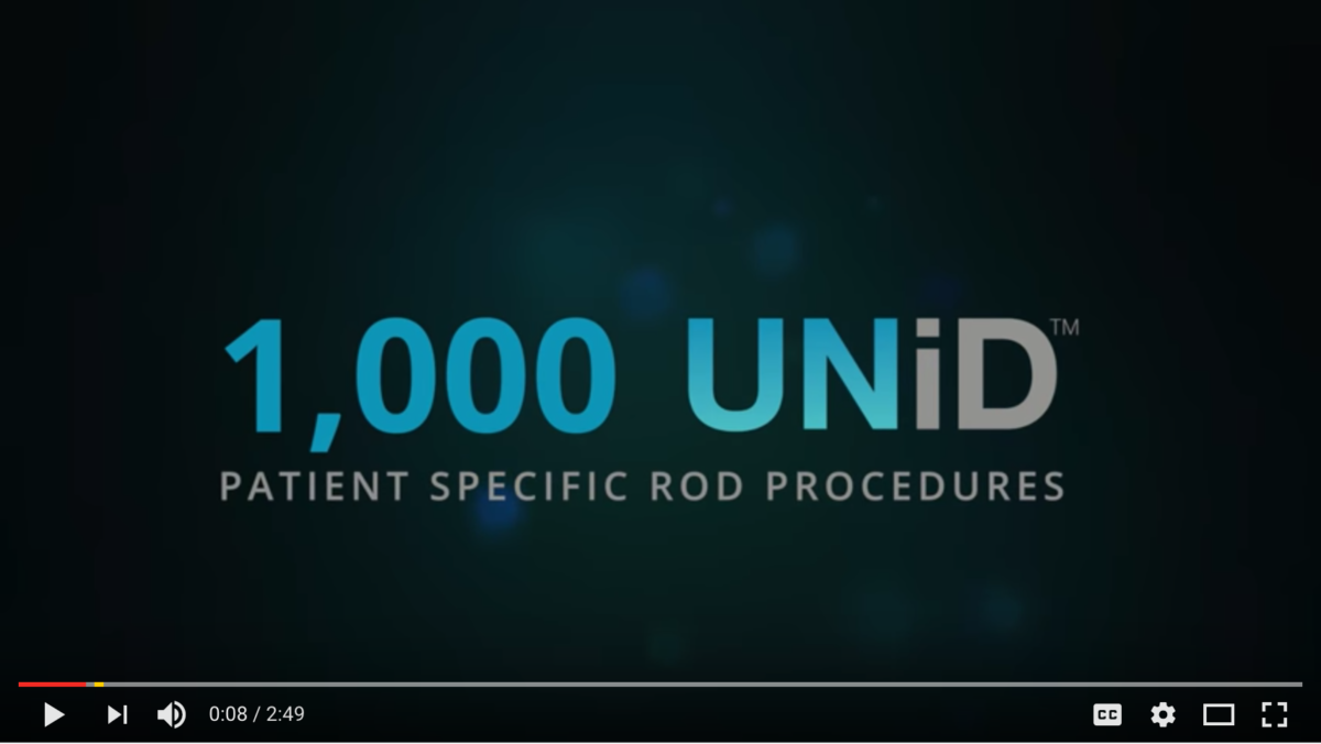
An advanced software tool helps surgeons to design patient-specific rods based on the simulation of the optimal sagittal alignment specific to each patient after measuring their spino-pelvic parameters.

2.ORDER
With one click, the order is placed with the UNiD Lab which processes the request and industrially produces the rods specifically for each patient.

3.EXECUTE YOUR PLAN

The UNiD patient-specific rods, designed to achieve the patient’s optimal sagittal alignment, are ready to be implanted. With UNiD, manual contouring of the rods during surgery becomes obsolete. Precious time in the operating room is saved and the surgeon can concentrate on executing the surgical plan for the patient’s best possible outcome.