Park City, UT

3 days / 6 sessions
Current Issues in Spine

February 2-4, 2017

AAEAAQAAAAAAAAJYAAAAJDRjNThjOTU3LWQwZDAtNDA2ZS1iMWM0LWY4NjZhZjJhMThiOA.png

September 7, 2016 OrthoSpineNews

September 07, 2016

CAMBRIDGE, Mass.–(BUSINESS WIRE)–InVivo Therapeutics Holdings Corp. (NVIV) today announced that Kristin Neff, Vice President of Clinical Operations & Project Management, is scheduled to present at the 55th International Spinal Cord Society (ISCoS) Annual Scientific Meeting to be held September 14-16, 2016 in Vienna, Austria. Ms. Neff was invited to present along with several other leaders in the field of spinal cord injury clinical research on September 14 during the symposium titled “Clinical Trials Update for 2016.” The symposium, organized by the Spinal Cord Outcomes Partnership Endeavor (SCOPE), is intended to provide an update on current clinical research to foster communication between researchers and clinicians on advancements and challenges in clinical research.

“I am excited to share our encouraging progress to date and partake in the discussion on the challenges of conducting clinical studies in the spinal cord injury patient population,” Ms. Neff said.

For more information regarding the meeting, visit:https://www.iscosmeetings.org/

About the Neuro-Spinal Scaffold™ Implant

Following acute spinal cord injury, surgical implantation of the biodegradable Neuro-Spinal Scaffold within the decompressed and debrided injury epicenter is intended to support appositional healing, thereby reducing post-traumatic cavity formation, sparing white matter, and allowing neural regeneration across the healed wound epicenter. The Neuro-Spinal Scaffold, an investigational device, has received a Humanitarian Use Device (HUD) designation and currently is being evaluated in the INSPIRE pivotal probable benefit study for the treatment of patients with complete (AIS A) traumatic acute spinal cord injury.

About InVivo Therapeutics

InVivo Therapeutics Holdings Corp. is a research and clinical-stage biomaterials and biotechnology company with a focus on treatment of spinal cord injuries. The company was founded in 2005 with proprietary technology co-invented by Robert Langer, Sc.D., Professor at Massachusetts Institute of Technology, and Joseph P. Vacanti, M.D., who then was at Boston Children’s Hospital and who now is affiliated with Massachusetts General Hospital. In 2011, the company earned the David S. Apple Award from the American Spinal Injury Association for its outstanding contribution to spinal cord injury medicine. In 2015, the company’s investigational Neuro-Spinal Scaffoldreceived the 2015 Becker’s Healthcare Spine Device Award. The publicly-traded company is headquartered in Cambridge, MA. For more details, visit www.invivotherapeutics.com.

Contacts

InVivo Therapeutics Holdings Corp.
Brian Luque, 617-863-5535
Investor Relations
bluque@invivotherapeutics.com


Drdk2-1200x718.jpg

September 1, 2016 OrthoSpineNews

By JOHN GAUDIOSI • August 28th, 2016

Dr. Robert Louis, a neurosurgeon at Hoag Memorial Hospital Presbyterian in Orange County, CA, is pitched some type of new technology, gadget or medication every day. He’s shown things so often that he developed an internal filter that automatically sets expectations a lot lower than the enthusiasm of the rep. But that all changed in October 2015.

That’s when Surgical Theater reps dropped by to showcase the Surgical Navigation Advanced Platform, or SNAP. Designed by former Israeli fighter pilots, the technology uses virtual reality to allow neurosurgeons to “fly” through a patient’s brain to get a better look at tumors, nerves, blood vessels and tissue prior to surgery. Before surgery, the patient’s brain is captured and recreated as a 3D model for Dr. Louis or his colleague Dr. Christopher Duma, neurosurgeon and director of Hoag’s Brain Tumor Program, to navigate.

Hoag is currently using an Oculus DK2, but the FDA recently cleared the consumer Oculus Rift for use and that will be deployed moving forward in all medical facilities, according to Jim Breidenstein, president and COO at Surgical Theater’s SNAP division.

Louis said prior to the introduction of this technology, he’d have to reference black-and-white 2D “slices” of the brain and then use his imagination (and 20 years of surgical experience) to map out the surgical procedure in his head before entering the Operating Room.

Since SNAP is registered with both Stealth, a technology Hoag uses, and Brainlab, that 3D model of the patient’s brain is used to track the tips of the instruments as the neurosurgeon navigates the brain. It works like a GPS inside the head, allowing doctors to track their instrument in real-time.

“Instead of looking at a 2D model, I can now see the tips of the instruments on the 3D Surgical Theater System on screen and compare that to what I’m seeing through the lens of the microscope,” said Louis, who is also director of Hoag’s Skull Base and Pituitary Tumor Program.


READ THE REST HERE


highresolution-1200x374.jpg

August 31, 2016 OrthoSpineNews

FREMONT, Calif., Aug. 31, 2016 /PRNewswire/ — Asterias Biotherapeutics, Inc. (NYSE MKT: AST), a biotechnology company focused on the emerging field of regenerative medicine, today announced that its Data Monitoring Committee (DMC) has reviewed the safety data from the initial cohort of three patients dosed with 2 million cells, and a subsequent five patients in the second cohort dosed with 10 million cells, and has cleared the company to now begin dosing a third cohort of 5-8 complete cervical injury patients (AIS-A patients) with the highest dose of 20 million cells.  Concurrently, the study is also proceeding with enrolling the first cohort of 5-8 sensory incomplete cervical spinal cord injury patients (AIS-B patients), each of whom will be administered 10 million cells.

The SCiStar study is an ongoing Phase 1/2a clinical trial funded in part by a $14.3 million grant from the California Institute for Regenerative Medicine (CIRM) and is designed to evaluate the safety and efficacy of escalating doses of AST-OPC1 in newly injured patients with sensory and motor complete cervical spinal cord injury (SCI), as well as newly injured patients with sensory incomplete SCI.  These patients are commonly referred to as AIS-A and AIS-B patients, respectively.  The results of the ongoing trial continue to support a positive safety profile for AST-OPC1.  There have been no serious or unexpected adverse events related to AST-OPC1, the administration procedure or the accompanying short course of low-dose immunosuppression in any of the patients treated with AST-OPC1, including five patients in an earlier Phase 1 trial with neurologically complete thoracic SCI.

“The positive safety data in the previous phase 1 study and in the ongoing phase 1/2a study gives us the confidence to now proceed to administration of 20 million cells, which based on our significant pre-clinical research is likely well within the dosing range where we would expect to see clinically meaningful improvement in these patients,” said Dr. Edward Wirth, Chief Medical Officer of Asterias Biotherapeutics.

The data set evaluating the efficacy results six months after implantation of 10 million AST-OPC1 cells in complete cervical spinal cord injury patients will be available in January 2017 and will focus on improvement in physical functioning of the upper extremities (fingers, hands and arms) of each treated patient utilizing scoring on the International Standards for Neurological Classification of Spinal Cord Injury (ISNCSCI scale).  The published literature and opinion leaders in the spinal cord injury field indicate that a two motor level improvement in functioning utilizing this validated scale is clinically meaningful for these patients and should be the key measure in the evaluation of new therapies.

“According to the published scientific literature as well as key opinion leaders that we speak to, a two motor level improvement in physical functioning on the ISNCSCI scale can translate into a significant improvement in quality of life, reduced need for daily care and increased ability to live independently for patients with cervical spinal cord injuries. We look forward to announcing the six month follow up data from this 10 million cell cohort in January 2017,” commented Steve Cartt, President & Chief Executive Officer.

About the SCiStar Trial

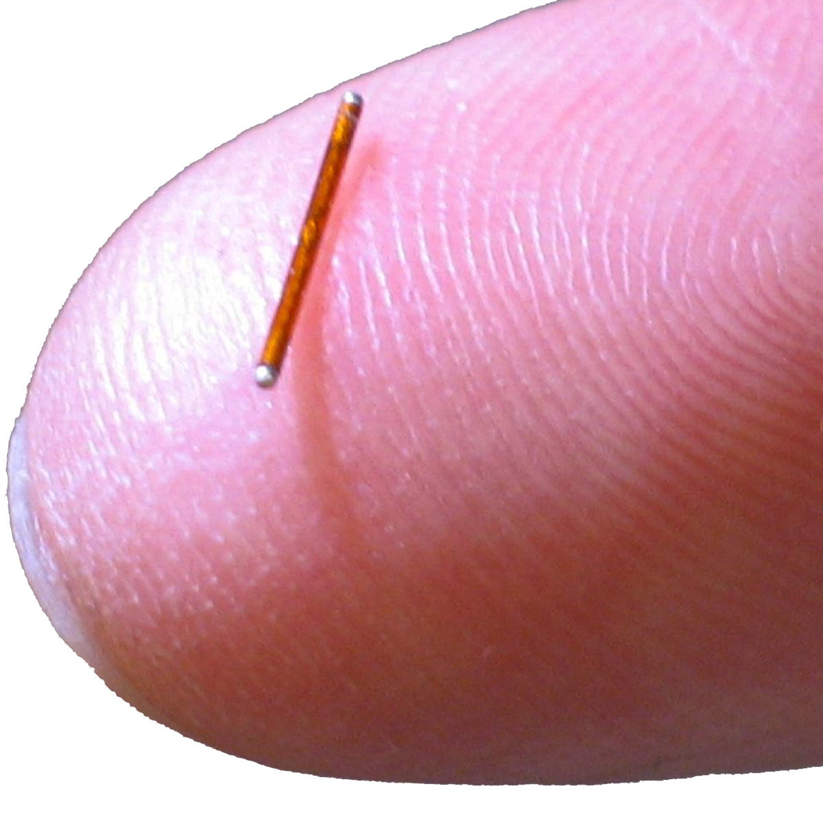
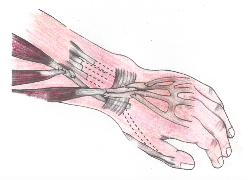
The SCiStar trial is testing three sequential escalating doses of AST-OPC1 administered at up to 20 million AST-OPC1 cells in as many as 35 patients with sub-acute, C-5 to C-7, motor complete (AIS-A or AIS-B) cervical SCI. These individuals have essentially lost all movement below their injury site and experience severe paralysis of the upper and lower limbs.  AIS-A patients have lost all motor and sensory function below their injury site, while AIS-B patients have lost all motor function but may retain some minimal sensory function below their injury site.  AST-OPC1 is being administered 14 to 30 days post-injury. Patients will be followed by neurological exams and imaging procedures to assess the safety and activity of the product. Additional information on the Phase 1/2a trial, including trial sites, can be found at www.clinicaltrials.gov, using Identifier NCT02302157, and at the SCiStar Study Website (www.scistar-study.com).

Asterias previously announced that it had been granted FDA clearance to expand patient enrollment in the Phase 1/2a clinical trial from 13 patients to up to 35 patients, based on the continued favorable safety profile observed in the ongoing clinical study. The Company believes that this change will increase the statistical confidence of the safety and efficacy readouts, reduce the risks of the AST-OPC1 program and position the product for potential accelerated regulatory approvals. Asterias has received a Strategic Partnerships Award grant from the California Institute for Regenerative Medicine, which provides $14.3 million of non-dilutive funding for the Phase 1/2a clinical trial and other product development activities for AST-OPC1.

More than 17,000 people sustain a spinal cord injury each year, but there are no FDA-approved therapeutics or devices that could potentially restore some function in individuals who have recently sustained a spinal cord injury.

About AST-OPC1

AST-OPC1, an oligodendrocyte progenitor population derived from human embryonic stem cells, has been shown in animals and in vitro to have three potentially reparative functions that address the complex pathologies observed at the injury site of a spinal cord injury. These activities of AST-OPC1 include production of neurotrophic factors, stimulation of vascularization, and induction of remyelination of denuded axons, all of which are critical for survival, regrowth and conduction of nerve impulses through axons at the injury site. In preclinical animal testing, AST-OPC1 administration led to remyelination of axons, improved hindlimb and forelimb locomotor function, dramatic reductions in injury-related cavitation and significant preservation of myelinated axons traversing the injury site.

In a previous Phase 1 clinical trial, five patients with neurologically complete, thoracic spinal cord injury were administered two million AST-OPC1 cells at the spinal cord injury site 7-14 days post-injury. They also received low levels of immunosuppression for the next 60 days.  Delivery of AST-OPC1 was successful in all five subjects with no serious adverse events associated with the administration of the cells, with AST-OPC1 itself, or the immunosuppressive regimen.  No evidence of rejection of AST-OPC1 was observed in detailed immune response monitoring of all patients.  In four of the five patients, serial MRI scans indicated that reduced spinal cord cavitation may have occurred.  Based on the results of this study, Asterias received approval from FDA to progress testing of AST-OPC1 to patients with complete cervical spine injuries, which represents the first targeted population for registration trials.

About Asterias Biotherapeutics

Asterias Biotherapeutics, Inc. is a leading biotechnology company in the emerging field of regenerative medicine. The company’s proprietary cell therapy programs are based on its immunotherapy and pluripotent stem cell platform technologies. Asterias is presently focused on advancing three clinical-stage programs which have the potential to address areas of very high unmet medical need in the fields of oncology and neurology. AST-OPC1 (oligodendrocyte progenitor cells) is currently in a Phase 1/2a dose escalation clinical trial in spinal cord injury. AST-VAC1 (antigen-presenting autologous dendritic cells) is being evaluated by Asterias for further development after demonstrating promise in a Phase 2 study in Acute Myeloid Leukemia (AML) and completing a successful end-of-Phase 2 meeting with the FDA. AST-VAC2 (antigen-presenting allogeneic dendritic cells) represents a second generation, allogeneic immunotherapy. The company’s research partner, Cancer Research UK, plans to begin a Phase 1/2 clinical trial of AST-VAC2 in non-small cell lung cancer in 2017. Additional information about Asterias can be found at www.asteriasbiotherapeutics.com.

FORWARD-LOOKING STATEMENTS

Statements pertaining to future financial and/or operating and/or clinical research results, future growth in research, technology, clinical development, and potential opportunities for Asterias, along with other statements about the future expectations, beliefs, goals, plans, or prospects expressed by management constitute forward-looking statements. Any statements that are not historical fact (including, but not limited to statements that contain words such as “will,” “believes,” “plans,” “anticipates,” “expects,” “estimates”) should also be considered to be forward-looking statements. Forward-looking statements involve risks and uncertainties, including, without limitation, risks inherent in the development and/or commercialization of potential products, uncertainty in the results of clinical trials or regulatory approvals, need and ability to obtain future capital, and maintenance of intellectual property rights. Actual results may differ materially from the results anticipated in these forward-looking statements and as such should be evaluated together with the many uncertainties that affect the businesses of Asterias, particularly those mentioned in the cautionary statements found in Asterias’ filings with the Securities and Exchange Commission. Asterias disclaims any intent or obligation to update these forward-looking statements.


SOURCE Asterias Biotherapeutics, Inc.

Related Links

http://www.asteriasbiotherapeutics.com


fingerStim-1-1200x1216.jpg

August 30, 2016 OrthoSpineNews

August 29, 2016

MIAMI–(BUSINESS WIRE)–Stimwave LLC, a medical device manufacturer and independent research institute headquartered in South Florida, has achieved long-term success for hundreds of pain patients during the early adaptor rollout of the world’s first miniature wireless pain relief system. Embraced by pain specialists seeking non-opioid treatments, the drug-free, breakthrough remedy offers new hope to the more than 400 million people worldwide who suffer from chronic pain.

“Every day in the U.S. an average of 120 people die as a result of drug overdose, more than from motor vehicle crashes, and another 6,700-plus are treated in emergency departments for the misuse or abuse of prescription pain medication,” said Stimwave Chairman and CEO Laura Tyler Perryman. “We believe that Stimwave’s Wireless Pain Relief® technology is not only a viable option to help millions of people who suffer from chronic pain, but a potential life-saver as the U.S. faces an epidemic of opioid addiction that in many cases is an unavoidable side effect of long-term use of addictive pain medication.”

Stimwave’s Wireless Pain Relief devices are 95 percent smaller than any other neuromodulation device on the market, so small, with a diameter of less than 1.5 millimeters, that they are simply implanted through a standard needle during an outpatient procedure and a minimally invasive receiver placement technique. Because they are wireless, with the power source discreetly worn outside the body, there is no need for the invasive “open surgery” required of earlier neuromodulation devices, reducing the risk of infection, pain at the site of a large battery, and other long-term complications of battery units and connectors that account for 85 percent of adverse events, according to registries. Further, this miniature wireless device is powered by a wearable antenna, easily integrated into clothing and requiring no sticky gels that could irritate the skin, and there are no “wires” protruding from the body.

“This is great news for pain specialists and chronic pain patients who previously did not have a minimally-invasive implant option available for pain,” said Dr. Sanjay Gupta, president of the American Pain Association and principal clinician at Atlantic Pain and Wellness Institute. “Our country is facing a horrible epidemic of drug overdose deaths. These wireless products provide an alternative to opioids, which is much needed in the armamentarium for effective pain control.”

Stimwave launched it’s FDA-cleared devices for the relief of chronic back and leg pain to a limited number of patients throughout 2015, and in March 2016 was granted FDA 510(k) for the relief of peripheral nervous system (PNS) pain, becoming the only neuromodulation device manufacturer cleared by the FDA to help reduce chronic neuropathic pain at most locations throughout the body, from back and leg pain addressed by spinal cord stimulation to PNS treatment for shoulder pain, wrist and elbow pain, knee pain, hip pain and more.

“The major issue with peripheral nerve stimulators in the past has always been the bulk and length of cables, connectors and pulse generators,” said Dr. Richard North, consultant and retired Professor of Neurosurgery at John Hopkins University School of Medicine. “A miniature wireless peripheral nerve stimulator will minimize the need for surgery in patients who already are suffering from pain. It has long been needed and now is finally a reality.”

Stimwave’s wireless device delivers small pulses of energy to specific nerves, triggering a reaction that enables the brain to remap pain pathways, thus providing pain relief. The Stimwave device contains no internal batteries or other toxic materials. The device is fixed in place by an anchor, so it stays “in line” with the body’s nerves, allowing a freedom of motion that is impossible with bulkier implanted devices.

The Stimwave portfolio also includes the only neuromodulation system approved for “full body” MRI examinations, meaning the device does not have to be removed for 3-Tesla MRI exams. This is a significant diagnostic breakthrough as it allows patients with pain to benefit from neuromodulation without restricting their access to MRI-assisted diagnoses, for current or future medical issues.

“Stimwave saved my life. Without this amazing product, I would still be in terrible pain, on medication, and having my health fail,” said Anthony Torres, a cargo crane operator severely injured in an industrial accident. [Please see accompanying Patient Profiles.] “The external wireless batteries are small, effective, and simple-to-use.

“The best thing about Stimwave is that it gave me relief from the pain and allowed me to get off the pain medications. I wanted to get off all the pain medications.”

Please visit www.stimwave.com for more information.

About Stimwave

Stimwave Technologies Incorporated is a privately held medical device company engaged in the development, manufacture, and commercialization of wirelessly powered, microtechnology neurostimulators, providing patients with a convenient, safe, minimally invasive, and highly cost-effective pain management solution that is easily incorporated into their daily lives. Stimwave’s goal is to evolve its patented, cutting-edge platform into the default for neuromodulation, increasing the accessibility for patients worldwide while lowering the economic impact of pain management.

Contacts

Glodow Nead Communications
Casey Shaughnessy, Sonia Sparks and Evan Nicholson, 415-394-6500
stimwavepr@glodownead.com


Tendon-Transfers_0.jpg

August 26, 2016 OrthoSpineNews

August 23, 2016

MAYWOOD, IL – A surgery for quadriplegics called tendon transfer can significantly improve hand and elbow function, but the procedure is greatly underused, according to an article in the journal Hand Clinics by Loyola Medicine hand surgeon Michael S. Bednar, MD, FAAOS.

In the procedure, muscles that still work are redirected to do the jobs of muscles that are paralyzed. Depending on the extent of the spinal cord injury, tendon transfers can enable a patient to grasp objects, pinch, open the hand and straighten the elbow. The patient can, for example, propel a wheelchair in the snow, use a fork without splints, grip a fishing pole, shake hands and perform daily activities such as dressing, bathing, toileting and transferring to and from a wheelchair.

“Although the long-term outcomes of these procedures are good, few patients eligible for these procedures actually have them performed,” Dr. Bednar wrote.

Dr. Bednar has performed tendon transfers on about 60 patients, and is among the most skilled and experienced surgeons in the country doing the procedure. Dr. Bednar is a professor in the department of orthopaedic surgery and rehabilitation at Loyola University Chicago Stritch School of Medicine.

When quadriplegics were asked what function they would most like restored, 75 percent said hand function, followed in order by bowel and bladder use (13 percent), walking (8 percent) and sexual performance (3 percent), according to an earlier study cited in Dr. Bednar’s article. However, only 14 percent of patients who are surgical candidates wind up getting tendon transfers, according to another previous study.

Patients who stand to benefit most from tendon transfers have spinal cord injuries in the C5-C8 cervical nerves in the lower neck. Patients must not have acute or chronic medical conditions such as infections, pressure sores, medical instability or spasticity.

“A good surgical candidate has functional goals, is motivated, understands benefits and limitations of surgery, demonstrates emotional and psychological stability/adjustment to disability and is committed to the post-operative rehabilitation process,” Dr. Bednar wrote.

Skeletal muscles come in pairs – one muscle to move the bone in one direction, another muscle to move it back. Muscles are connected to bones by tendons. The bone moves when the brain sends a signal down a nerve telling the muscle to contract.

In many cases, more than one muscle performs the same function. So in a tendon transfer, the surgeon shifts the tendon of one of the spare muscles to a new location. For example, the surgeon may detach one of the working elbow muscles (the brachioradialis) and reattach it to a nonworking muscle that flexes the thumb (the flexor pollicis longus).

The number of functioning muscles a patient has will determine what tendon transfers the surgeon will perform. The more working muscles available for transfer, the more functions can be restored.

Tendon transfers typically involve two surgeries on each arm, performed three months apart. Arms are done one at a time. During rehabilitation, patients learn how to use the transferred muscles.

Tendon transfers temporarily reduce hand and elbow function during recovery and rehabilitation. Tendon transfers also do not restore full function. But while pinch strength and grasp strength after rehabilitation are not as high as in a normal hand, they are high enough to perform most activities of daily living.

Among the reasons so few patients get tendon transfers are lack of communication among rehabilitation specialists, physicians and surgeons, poor access to care and lack of awareness. The greatest barrier appears to be a lack of coordinated collaboration among specialists, Dr. Bednar wrote.

Dr. Bednar concluded: “Continued education of patients with tetraplegia, their caregivers and the rehabilitation community will hopefully increase utilization of these effective tendon transfer procedures.” (Tetraplegia is another term for quadriplegia.)

Dr. Bednar’s paper is titled “Tendon Transfers for Tetraplegia.”

– Loyola University Health System


download.jpe

August 25, 2016 OrthoSpineNews

SAN DIEGO – A robot created to help surgeons during complicated procedures is bringing new hope to young patients who suffer neurological disorders.

Rady Children’s Hospital is among the few in the country using the technology.

The Rosa Robot has two main parts – a computer brain and a robotic arm.  It allows surgeons to create a 3D map of a patient’s brain.

“I think for the future of epilepsy centers and any center that wants to treat epilepsy in the future, this is a vital importance. Rady Children’s is one of the first five hospitals in the country to get this robotic technology to enable us to do that and its important in leading the field for it and continue to treating epilepsy in the best way possible,” said Dr. David Gonda, director of surgical epilepsy, Rady Children’s Hospital.

Rosa is a robotized surgical assistant – the most advanced of its kind.

It’s one of a few being used across the country and six months ago it arrived here at Rady Children’s hospital.

So, far its helped about twenty patients.

Through the robot, doctors are able to look at a 3D image from any angle and at any depth.


READ THE REST HERE


nerve-regeneration-device-625.jpg

August 24, 2016 OrthoSpineNews

By: Norbert Sparrow | PLASTEC Minneapolis

One of the rightly touted breakthroughs of 3D printing in the medical space is the technology’s capability of producing custom orthopedic devices that precisely match specific patient anatomies. University of Minnesota mechanical engineering professor Michael McAlpine wants to take that a step further by integrating customized functionality into the 3D-printed device.

“The big value in 3D printing is customization not just of the geometry but also the functionality,” McAlpine told PlasticsToday. “That means building in function from various materials that also may be specific to what the patient needs.” He and fellow researchers have taken a big step in that direction with a 3D-printed silicone guide embedded with proteins to enable nerve regeneration. The technique has been shown to be successful in regenerating the sciatic nerve in rats.

Nerve regeneration in and of itself is a complex process, and that is compounded by the y-shaped geometry of the sciatic nerve, which branches out as it travels down the leg. “A damaged sciatic nerve is difficult, if not impossible, to repair,” says McAlpine. Through the use of a 3D scanner and custom designed 3D printer that allows integration of multiple materials, including plastics, cells and functional materials such as electronic and semiconducting nanoparticles and hydrogels, on the same platform, McAlpine’s team was able to print custom nerve guides that promote both motor and sensory nerve regeneration.

READ THE REST HERE