Park City, UT

3 days / 6 sessions
Current Issues in Spine

February 2-4, 2017

npt1.jpg

September 8, 2016 OrthoSpineNews

September 08, 2016

DURHAM, N.C.–(BUSINESS WIRE)–Recent research has shed light on the predictability of fractures that fail to heal, known as nonunions. All nonunions are a function of severity, location and disease comorbidity and while risk factor interaction is complex, a new study reports that it may become possible to predict nonunion based on the patient-specific presentation of risk factors. The findings are published in the September 7, 2016 issue of JAMA Surgery available at http://archsurg.jamanetwork.com/article.aspx?articleid=2547685.

The study found that in 2011, among 309,330 fractures in 18 bones, the overall nonunion rate was 4.93%. However, higher nonunion risk was associated with severe fractures, high body mass index, smoking and alcoholism. While females had more fractures, males were more prone to nonunion. In addition, the risk of nonunion increased for patients who used certain medications including antibiotics, anticoagulants, and opioids, as well as for patients who had diseases such as obesity, osteoarthritis and osteoporosis.

“We hypothesized that the interplay between a patient’s physiological risk factors and fracture characteristics increased the risk of fracture nonunion,” said Dr. R. Grant Steen, Manager of Medical Affairs, Bioventus. “We now believe clinicians can use this information to describe the epidemiology of fracture nonunion in adult patients.”

The study was funded by Bioventus and used fracture patients from a health plan database. Patients with fracture were identified and continuous enrollment in the database was required for 12 months after fracture, to allow sufficient time to capture a nonunion diagnosis.

Authors of this study include Robert Zura, MD, LSU Health Science Center, New Orleans, Ze Xiong, MS, Dept. of Statistics, North Carolina State University, Thomas Einhorn, MD, NYU Langone Medical Center, J. Tracy Watson, MD Saint Louis University School of Medicine, Robert F. Ostrum, MD University of North Carolina, Michael J. Payson, MD, Wright State University, Gregory J. Della Rocca, MD, PhD, University of Missouri, Samir Mehta, MD, Hospital of the University of Pennsylvania, Todd McKinley, MD, Indiana University, Zhe Wang, MS, Dept. of Statistics, North Carolina State University and R. Grant Steen, PhD, Manager of Medical Affairs, Bioventus.

About Bioventus

Bioventus is an orthobiologics company that delivers clinically proven, cost-effective products that help people heal quickly and safely. Its mission is to make a difference by helping patients resume and enjoy active lives. Bioventus has two product portfolios for orthobiologics, Bioventus Active Healing Therapies and Bioventus Surgical that make it a global leader in active orthopaedic healing. Built on a commitment to high quality standards, evidence-based medicine and strong ethical behavior, Bioventus is a trusted partner for physicians worldwide.

For more information, visit www.BioventusGlobal.com and follow the company on Twitter @Bioventusglobal

Bioventus and the Bioventus logo are registered trademarks of Bioventus LLC.

Contacts

Bioventus
Thomas Hill, 919-474-6715
thomas.hill@bioventusglobal.com


AAEAAQAAAAAAAAJYAAAAJDRjNThjOTU3LWQwZDAtNDA2ZS1iMWM0LWY4NjZhZjJhMThiOA.png

September 7, 2016 OrthoSpineNews

September 07, 2016

CAMBRIDGE, Mass.–(BUSINESS WIRE)–InVivo Therapeutics Holdings Corp. (NVIV) today announced that Kristin Neff, Vice President of Clinical Operations & Project Management, is scheduled to present at the 55th International Spinal Cord Society (ISCoS) Annual Scientific Meeting to be held September 14-16, 2016 in Vienna, Austria. Ms. Neff was invited to present along with several other leaders in the field of spinal cord injury clinical research on September 14 during the symposium titled “Clinical Trials Update for 2016.” The symposium, organized by the Spinal Cord Outcomes Partnership Endeavor (SCOPE), is intended to provide an update on current clinical research to foster communication between researchers and clinicians on advancements and challenges in clinical research.

“I am excited to share our encouraging progress to date and partake in the discussion on the challenges of conducting clinical studies in the spinal cord injury patient population,” Ms. Neff said.

For more information regarding the meeting, visit:https://www.iscosmeetings.org/

About the Neuro-Spinal Scaffold™ Implant

Following acute spinal cord injury, surgical implantation of the biodegradable Neuro-Spinal Scaffold within the decompressed and debrided injury epicenter is intended to support appositional healing, thereby reducing post-traumatic cavity formation, sparing white matter, and allowing neural regeneration across the healed wound epicenter. The Neuro-Spinal Scaffold, an investigational device, has received a Humanitarian Use Device (HUD) designation and currently is being evaluated in the INSPIRE pivotal probable benefit study for the treatment of patients with complete (AIS A) traumatic acute spinal cord injury.

About InVivo Therapeutics

InVivo Therapeutics Holdings Corp. is a research and clinical-stage biomaterials and biotechnology company with a focus on treatment of spinal cord injuries. The company was founded in 2005 with proprietary technology co-invented by Robert Langer, Sc.D., Professor at Massachusetts Institute of Technology, and Joseph P. Vacanti, M.D., who then was at Boston Children’s Hospital and who now is affiliated with Massachusetts General Hospital. In 2011, the company earned the David S. Apple Award from the American Spinal Injury Association for its outstanding contribution to spinal cord injury medicine. In 2015, the company’s investigational Neuro-Spinal Scaffoldreceived the 2015 Becker’s Healthcare Spine Device Award. The publicly-traded company is headquartered in Cambridge, MA. For more details, visit www.invivotherapeutics.com.

Contacts

InVivo Therapeutics Holdings Corp.
Brian Luque, 617-863-5535
Investor Relations
bluque@invivotherapeutics.com


AZ2ZgMjz0LFGHCPn4xMDoxOjA4MTsiGN.620x360_q85_upscale.jpg

September 7, 2016 OrthoSpineNews

September 07, 2016

PLYMOUTH, Minn.–(BUSINESS WIRE)–Rotation Medical Inc., a medical device company focused on developing new technologies to treat rotator cuff disease, today announced that study results published in the current issue of Muscle, Ligaments and Tendons Journal showed that the company’s collagen-based bioinductive implant induced new tissue formation in all study patients with rotator cuff tears. The Rotation Medical rotator cuff system is a novel, implant-based solution for rotator cuff repair and a new alternative to traditional surgical repair.

The study assessed the ability of Rotation Medical’s bioinductive implant to induce new tissue formation and limit tear progression when placed on the bursal surface of partial thickness cuff tears. The implant induced significant new tissue formation in all 13 patients by three months (mean increase in tendon thickness 2.2 ± 0.26 mm), and the tissue matured over time and became radiologically indistinguishable from the underlying tendon. No tear progression was observed on MRI in any of the patients during the 24-month post-operative period. All patients’ Constant and American Shoulder and Elbow Society clinical scores improved significantly over time.1

“Partial-thickness rotator cuff tears frequently enlarge due to increased local strain and often progress to full-thickness tears,” said Dr. Desmond John Bokor, lead study investigator and associate professor in the Department of Orthopaedic Surgery at Macquarie University in Australia. “The results of this study demonstrate the ability of the bioinductive implant to induce new tendon-like tissue, enabling partial-thickness rotator cuff tears to decrease in size and in most cases disappear. The ability to heal partial-thickness rotator cuff defects, and thus prevent tear propagation and progressive tendon degeneration, represents a novel interventional treatment paradigm for these lesions.”

Rotator cuff damage is the most common source of shoulder pain, affecting more than 4 million people annually in the U.S. Traditional approaches to treating degenerate or torn rotator cuffs often do not address the poor quality of the underlying tendon tissue, and a significant number of these tendons, after standard treatment, either degenerate further and/or re-tear. Cleared by the U.S. Food and Drug Administration in March 2014, the Rotation Medical bioinductive implant is designed to address this limitation by inducing new tissue growth at the site of implantation, resulting in increased tendon thickness and healing of tendon defects with new tissue growth. The collagen-based implant is about the size of a postage stamp and it is part of the Rotation Medical rotator cuff system, which also includes disposable instruments that allow the arthroscopic procedure to be performed easily and quickly.

“This study is further evidence that the Rotation Medical rotator cuff system has the potential to transform the treatment of rotator cuff disease,” said Martha Shadan, president and CEO of Rotation Medical. “Our bioinductive implant addresses both the biomechanics and biology required to heal a rotator cuff tendon tear, preventing rotator cuff tears from becoming larger over time, reducing the incidence of re-tears and, in some cases, shortening patient recovery time.”

The study, “Evidence of healing of partial-thickness rotator cuff tears following arthroscopic augmentation with a collagen implant: a 2-year MRI follow-up,” adds to the growing body of literature supporting the use of the bioinductive implant as a novel treatment for rotator cuff partial thickness tears. Additional publications and information about the Rotation Medical rotator cuff system are available on the company’s website.

About the Study

The implant was inserted via arthroscopic surgery. A total of 13 patients with intermediate (3-6 mm) to high-grade (>6 mm) partial thickness cuff tears completed two years of follow-up. At three, six, 12, and 24 months postoperatively, tendon thickness, defect size and quality were evaluated using magnetic resonance imaging (MRI), and clinical outcomes were assessed using the Constant and American Shoulder and Elbow Society scores. The partial-thickness cuff tears showed consistent filling of the defects, with complete healing in seven patients at 12 months, and a progressive improvement in tendon quality in the remaining patients. No tear progression was observed by MRI in any of the patients at 24 months. All clinical scores improved significantly over time. At 24 months, 12 of 13 patients (92 percent) had satisfactory or better results.

About Rotation Medical

Rotation Medical Inc. was founded in 2009 and is committed to improving the treatment of rotator cuff disease with the Rotation Medical rotator cuff system, a breakthrough technology that has the potential to prevent rotator cuff disease progression and reduce re-tears by inducing the growth of new tendinous tissue. The company is privately held and funded by New Enterprise Associates (NEA), Life Sciences Partners (LSP) and Pappas Ventures. For more information, visit www.rotationmedical.com.

________________________
1 Bokor DJ, Sonnabend D, Deady L, Cass B, Young A, Van Kampen C, Arnoczky S. Evidence of healing of partial-thickness rotator cuff tears following arthroscopic augmentation with a collagen implant: a 2-year MRI follow-up. Muscle Ligaments Tendons J. 2016 May 19;6(1):16-25. doi: 10.11138/mltj/2016.6.1.016.

Contacts

Merryman Communications
Joni Ramirez, 323-532-0746
joni@merrymancommunications.com


srep01408-f1-1.jpg

September 6, 2016 OrthoSpineNews

September 2, 2016 – Ajayan Group/Rice University

The Rice lab of materials scientist Pulickel Ajayan and colleagues in Texas, Brazil and India used spark plasma sintering to weld flakes of graphene oxide into porous solids that compare favorably with the mechanical properties and biocompatibility of titanium, a standard bone-replacement material.

The discovery is the subject of a paper in Advanced Materials.

The researchers believe their technique will give them the ability to create highly complex shapes out of graphene in minutes using graphite molds, which they believe would be easier to process than specialty metals.

“We started thinking about this for bone implants because graphene is one of the most intriguing materials with many possibilities and it’s generally biocompatible,” said Rice postdoctoral research associate Chandra Sekhar Tiwary, co-lead author of the paper with Dibyendu Chakravarty of the International Advanced Research Center for Powder Metallurgy and New Materials in Hyderabad, India. “Four things are important: its mechanical properties, density, porosity and biocompatibility.”

Tiwary said spark plasma sintering is being used in industry to make complex parts, generally with ceramics. “The technique uses a high pulse current that welds the flakes together instantly. You only need high voltage, not high pressure or temperatures,” he said. The material they made is nearly 50 percent porous, with a density half that of graphite and a quarter of titanium metal. But it has enough compressive strength — 40 megapascals — to qualify it for bone implants, he said. The strength of the bonds between sheets keeps it from disintegrating in water.

The researchers controlled the density of the material by altering the voltage that delivers the highly localized blast of heat that makes the nanoscale welds. Though the experiments were carried out at room temperature, the researchers made graphene solids of various density by raising these sintering temperatures from 200 to 400 degrees Celsius. Samples made at local temperatures of 300 C proved best, Tiwary said. “The nice thing about two-dimensional materials is that they give you a lot of surface area to connect. With graphene, you just need to overcome a small activation barrier to make very strong welds,” he said.


READ THE REST HERE


September 1, 2016 OrthoSpineNews

MARIETTA, Ga., Sept. 1, 2016 /PRNewswire/ — MiMedx Group, Inc. (NASDAQ: MDXG), the leading regenerative medicine company utilizing human amniotic tissue and patent-protected processes to develop and market advanced products and therapies for the Wound Care, Surgical, Orthopedic, Spine, Sports Medicine, Ophthalmic, and Dental sectors of healthcare, announced today its plans for the nationwide launch of AmnioFill™, the first product in the MiMedx placental collagen matrix product family to be commercially launched.

Physicians are in need of a product to treat larger acute and chronic wounds encountered in the surgical setting. Slated for nationwide release later this month, AmnioFill is being offered in multiple sizes and configurations to address this and other surgical needs.

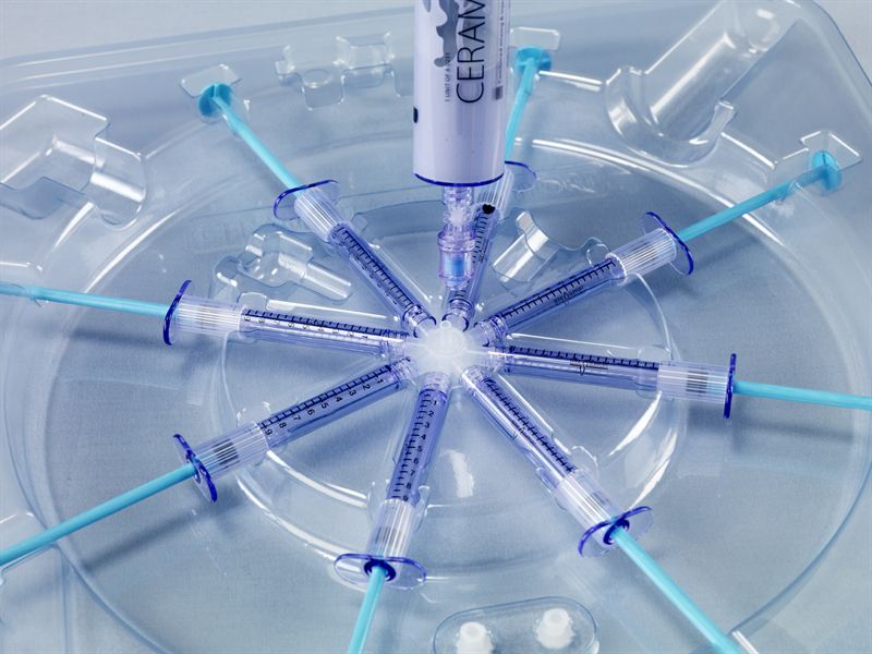
AmnioFill is a collagenous matrix derived from the placenta and comprised of placental extracellular matrix (ECM) tissue. AmnioFill is a tissue allograft containing ECM proteins, growth factors, cytokines and other specialty proteins present in placental tissue. Over 226 growth factors, cytokines and chemokines, including important modulators of inflammation and factors critically important in wound healing, are contained in the AmnioFill placental tissue. MiMedx employs terminal sterilization in addition to aseptic processing techniques in its proprietary processing methodology to enhance the safety of AmnioFill and its other amniotic and placental products.

Parker H. “Pete” Petit, Chairman and CEO, said, “AmnioFill will be a great addition to our product lines, addressing the needs of both Wound Care and Surgical markets. For example, we expect that AmnioFill will be an ideal solution for physicians in the treatment of dehisced surgical wounds and other deep complex and hard-to-heal surgical wounds that require a connective tissue matrix to replace or supplement damaged or inadequate integumental tissue.”

Christopher M. Cashman, Executive Vice President and Chief Commercialization Officer, commented, “The historical costs to treat these types of wounds are significant, and the quality of life issues of non-resolved wounds of this nature can be devastating.  There were 53 million outpatient procedures performed in the United States in 2010. Despite advances in preoperative care, the rate of surgical wound dehiscence has not decreased in recent years with 1% to 3% of patients experiencing wound dehiscence. For example, breast reconstruction incisional dehiscence rates range from 10% to 15% in a setting with radiation therapy and abdominal wall surgical dehiscences have a mortality rate as high as 45%.”

Bill Taylor, President and COO, stated, “Our published scientific studies have demonstrated that our dehydrated Human Amnion/Chorion Membrane (dHACM) allografts cause stem cells to migrate and proliferate. Moreover, these scientific studies also demonstrated our dHACM allografts promote angiogenesis. Multiple clinical studies have confirmed that stem cell migration, proliferation and recruitment as well as angiogenesis are essential in wound healing.”

Cashman added, “AmnioFill is designed to provide a scaffold for recruited cells to attach, populate and proliferate. The placental tissues in the scaffold should modulate the activity of the recruited cells to generate new tissue for these larger acute and chronic surgical wounds. When used earlier in the treatment of these complex wounds, we believe AmnioFill becomes an even more cost effective approach as a step therapy for wound closure.”

About MiMedx

MiMedx® is an integrated developer, processor and marketer of patent protected and proprietary regenerative biomaterial products and bioimplants processed from human amniotic membrane and other birth tissues and human skin and bone.  “Innovations in Regenerative Biomaterials” is the framework behind our mission to give physicians products and tissues to help the body heal itself.  The MiMedx allograft product families include our: dHACM family with AmnioFix®, EpiFix® and EpiBurn® brands; Amniotic Fluid family with OrthoFlo brand; Umbilical family with EpiCord™ and AmnioCord™ brands; Placental Collagen family with CollaFix™ and AmnioFill™ brands; Bone family with Physio® brand; and Skin family with AlloBurn™ brand. AmnioFix,  EpiFix, and EpiBurn are our tissue technologies processed from human amniotic membrane; OrthoFlo is an amniotic fluid derived allograft;  EpiCord™ and AmnioCord™ are derived from the umbilical cord; Physio is a unique bone grafting material comprised of 100% bone tissue with no added carrier; AlloBurn is a skin product derived from human skin designed for the treatment of burns; and  CollaFix, our next brand we plan to commercialize, is our collagen fiber technology, developed with our patented cross-linking polymers, designed to mimic the natural composition, structure and mechanical properties of musculoskeletal tissues in order to augment their repair.

We process the human amniotic membrane utilizing our proprietary PURION® Process, to produce a safe and effective implant. MiMedx proprietary processing methodology employs aseptic processing techniques in addition to terminal sterilization.  MiMedx is the leading supplier of amniotic tissue, having supplied over 700,000 allografts to date for application in the Wound Care, Burn, Surgical, Orthopedic, Spine, Sports Medicine, Ophthalmic and Dental sectors of healthcare.

Safe Harbor Statement

This press release includes statements that look forward in time or that express management’s beliefs, expectations or hopes. Such statements are forward-looking statements within the meaning of the Private Securities Litigation Reform Act of 1995. These statements include, but are not limited to the Company’s belief that AmnioFill will be a great addition to its product lines, addressing the needs of both wound care and surgical markets; that AmnioFill will be an ideal solution for physicians in the treatment of dehisced surgical wounds and other deep complex and hard-to-heal surgical wounds that require a connective tissue matrix to replace or supplement damages or inadequate integumental tissue; and that, when used earlier in the treatment of complex wounds, AmnioFill becomes an even more cost effective approach as a step therapy for wound closure. Among the risks and uncertainties that could cause actual results to differ materially from those indicated by such forward-looking statements include that demand for, and acceptance of, any new product by the medical community may not be as expected; factors such as third party reimbursement may impact physician use of product; AmnioFill may not be used as anticipated or perform as anticipated in the clinical setting; AmnioFill may  not be as cost effective as anticipated, and the risk factors detailed from time to time in the Company’s periodic Securities and Exchange Commission filings, including, without limitation, its 10-K filing for the fiscal year ended December 31, 2015 and its most recent 10Q filing. By making these forward-looking statements, the Company does not undertake to update them in any manner except as may be required by the Company’s disclosure obligations in filings it makes with the Securities and Exchange Commission under the federal securities laws.

SOURCE MiMedx Group, Inc.

Related Links

http://www.mimedx.com


bc93a3c1a37b0541_800x800ar.jpg

September 1, 2016 OrthoSpineNews

Lund, Sweden, 1 September 2016 – BONESUPPORT AB, an emerging leader in innovative injectable bioresorbable bone graft substitute products to treat bone voids caused by trauma, infection, disease or related surgery, today announced the publication of a paper in The Bone and Joint Journal: Single-stage treatment of chronic osteomyelitis with a new absorbable gentamicin-loaded, calcium sulphate/hydroxyapatite biocomposite – A prospective series of 100 cases. McNally et al, The Bone and Joint Journal, 2016, Vol. 98-B, No. 9, p1289-96.

The paper provides 12-34 month follow up data from the first 100 patients in a prospective study evaluating CERAMENT G for dead space (void) management in patients with chronic osteomyelitis (bone infection) using a single stage surgical procedure. These data showed that this approach, augmented by the use of CERAMENT G, was highly effective, delivering a 96% prevention of infection recurrence rate, a 3.0% fracture rate and a total wound leakage rate of 6.0%.  This is significantly lower than published results with alternative bone graft substitutes that deliver antibiotics locally.

These results highlight the essential properties of CERAMENT G in the management of chronic osteomyelitis. The very encouraging infection recurrence prevention rate is supported by CERAMENT G’s attractive local delivery properties, which enable it to provide an initial targeted ultra-high concentration of gentamicin into the bone defect and then a longer sustainable dose above the minimal inhibitory concentration (MIC) of the bacteria that initially caused the osteomyelitis. This unique antibiotic-eluting profile helps protect the bone healing process and promote bone remodeling.

The bone healing and bone remodeling properties of CERAMENT G, when combined with gentamicin, make it an ideal solution for dead space management in patients with chronic osteomyelitis. It is able to fill the void completely due to its injectibility and to provide initial structural stability due to its self-setting properties.

The use of CERAMENT G to deliver gentamicin locally could play an important role in improving antibiotic stewardship in hospitals by increasing compliance and reducing the need for patients with chronic osteomyelitis to receive long term systemic antibiotics.

Mr Martin McNally, Consultant Bone Infection and Limb Reconstruction Surgeon at Oxford University Hospitals (Oxford, UK) and lead author of the paper said, “The results that we have achieved with the single stage surgical procedure using CERAMENT G for the dead space management of patients with chronic osteomyelitis are a significant improvement on past experience. These results reflect CERAMENT G’s unique local antibiotic delivery profile and its attractive bone remodelling capabilities. We are increasingly using CERAMENT G in the treatment of patients with chronic osteomyelitis and infected fractures. It allows a more patient-friendly treatment, preventing repeated operations and recurrent infections. We expect it to become the mainstay of our dead space management, given the major clinical and health economic benefits that it supports.”

The paper covers the first 100 patients in a prospective cohort study utilising CERAMENT G for dead space management in a single stage surgical procedure for chronic osteomyelitis. The mean duration of chronic osteomyelitis in this patient group was 10.4 years (0.5 to 68 years). All surgeries were performed by two surgeons and were completed in a single operative session. All patients were given similar systemic antibiotic therapy and rehabilitation. Patients were followed up for at least 12 months (mean 19.5 months, range 12 -34 months) with infection recurrence, fracture rate and wound leakage rate as the primary outcome measure. The study showed that the single stage surgical procedure with CERAMENT G, was highly effective delivering a 96% infection recurrence prevention rate, a 3.0% fracture rate and a total wound leakage rate of 6.0%.

Richard Davies, CEO of BONESUPPORT said, “The results that have been published today highlight the clear clinical benefits  with CERAMENT G’s ability to deliver sustained bactericidal levels of gentamicin locally to support the eradication of underlying infections in patients with chronic osteomyelitis. By using CERAMENT G in a single stage procedure to help patients with chronic osteomyelitis return to a normal life, we can deliver significant health economic benefits to payors who are struggling to contain the significant and growing costs of treating severely debilitating bone infection.”

Reference

McNally et al, The Bone and Joint Journal, 2016, Vol. 98-B, No. 9, p1289-96.

Notes to Editor

About BONESUPPORT™

BONESUPPORT has developed CERAMENT an innovative range of radiopaque injectable bone graft substitute products that have a proven ability to heal defects by remodeling to host bone in six to twelve months. Our products are effective in treating patients with fractures and bone voids caused by trauma, infection, disease or related surgery. Our lead product, CERAMENT BVF addresses important issues facing health care providers, such as avoiding hospital readmissions and revision surgery that result from failed bone healing and infection caused by residual bone voids. CERAMENT BVF is commercially available in the U.S., EU, SE Asia and the Middle East.

CERAMENT BVF’s distinctive properties as a drug eluting material have been validated in clinical practice by CERAMENT G and CERAMENT V, the first CE-marked injectable antibiotic eluting bone graft substitutes. These products provide local sustained delivery of gentamicin and vancomycin, respectively. The local delivery feature enables an initial high concentration of antibiotics to the bone defect and then a longer sustainable dose above the minimal inhibitory concentration (MIC) to protect bone healing and promote bone remodeling.

CERAMENT G and V have demonstrated good results in patients with problematic bone infections including osteomyelitis. They are also used prophylactically in patients who are at risk for developing infection. CERAMENT G and CERAMENT V are available in the EU.

BONESUPPORT was founded in 1999 by Prof. Lars Lidgren, an internationally respected scientist who has been the President of various musculoskeletal societies. BONESUPPORT’s mission is to bring people with bone and joint diseases back to an active life. The Company is based in Lund, Sweden with subsidiary locations in the U.S. and Germany. www.bonesupport.com

BONESUPPORT™ is a registered trademark.


Contact Information


Citigate Dewe Rogerson

David Dible, Andrea Bici

+44 (0)20 7282 2949/1050

bonesupport@citigatedr.co.uk


BONESUPPORT AB 

Richard Davies, CEO

Phone +46 46 286 53 59

Richard.Davies@bonesupport.com


MM09116LOGO-3-1200x339.jpe

August 30, 2016 OrthoSpineNews

ENGLEWOOD, Colo., Aug. 30, 2016 /PRNewswire/ — Ampio Pharmaceuticals, Inc. (NYSE MKT: AMPE) announced today publication in the peer-reviewed journal Biochemistry and Biophysics Reports that further describes the modes of action (MOA) of Ampion in the treatment of Osteoarthritis. The publication is titled:

“The low molecular weight fraction of human serum albumin upregulates COX2, prostaglandin E2, and prostaglandin D2 under inflammatory conditions in osteoarthritic knee synovial fibroblasts.”  Elizabeth D. Frederick, Ph.D.; Melissa A. Hausburg, Ph.D.;Gregory W. Thomas, BS; Leonard Rael, MS; Edward Brody, MD; David Bar-Or, MD and can be retrieved athttp://dx.doi.org/10.1016/j.bbrep.2016.08.015

Dr. David Bar-Or, Ampio’s Chief Science Officer, noted, “This manuscript reports that in the presence of either IL-1β or TNFα, LMWF-5A (Ampion™) increased the expression of both COX2 mRNA and protein, and this increase was significant compared to that observed with IL-1β- or TNFα-stimulated, saline-treated cells. LMWF-5A appears to trigger increased anti-inflammatory PG signaling, and this may be a primary component of its therapeutic mode of action in the treatment of OAK.” In simple words, these findings indicate that the mechanism of action of Ampion™ is through the production of beneficial prostaglandins by synoviocytes for both the resolution of inflammation and tissue regeneration.

About Osteoarthritis

Osteoarthritis (OA) is a progressive disorder of the joints involving degradation of the intra-articular cartilage, joint lining, ligaments, and bone. The incidence of developing osteoarthritis of the knee over a lifetime is approximately 45%. As this disease is associated with age, obesity, and diabetes this number will continue to grow. Certain risk factors in conjunction with natural wear and tear lead to the breakdown of cartilage. Osteoarthritis is caused by inflammation of the soft tissue and bony structures of the joint, which worsens over time and leads to progressive thinning of articular cartilage. Other symptoms include narrowing of the joint space, synovial membrane thickening, osteophyte formation and increased density of subchondral bone.

About Ampio Pharmaceuticals

Ampio Pharmaceuticals, Inc. is a development stage biopharmaceutical company primarily focused on the development of therapies to treat prevalent inflammatory conditions for which there are limited treatment options. We are developing compounds that decrease inflammation by (i) inhibiting specific pro-inflammatory compounds by affecting specific pathways at the protein expression and at the transcription level; (ii) activating specific phosphatase or depletion of the available phosphate needed for the inflammation process, and (iii) decreasing vascular permeability.

Forward Looking Statements

Ampio’s statements in this press release that are not historical fact, and that relate to future plans or events, are forward-looking statements within the meaning of the Private Securities Litigation Reform Act of 1995. Forward-looking statements can be identified by the use of words such as “believe,” “expect,” “plan,” “anticipate,” and similar expressions. These forward-looking statements include statements regarding Ampio’s scientific presentations on our Ampion™ product. The risks and uncertainties involved include those detailed from time to time in Ampio’s filings with the Securities and Exchange Commission, including without limitation, under Ampio’s Annual Report on Form 10-K and Quarterly Reports on Form 10-Q. Ampio undertakes no obligation to revise or update these forward-looking statements, whether as a result of new information, future events or otherwise.

Investor Contact:
Gregory A. Gould
Ampio Pharmaceuticals, Inc.
Phone: (720) 437-6500
E-mail: info@ampiopharma.com

(1) Burden of Major Musculoskeletal Conditions; Woolf, Pfleger; Bulletin of the World Health Organization

Logo – http://photos.prnewswire.com/prnh/20120516/MM09116LOGO 

SOURCE Ampio Pharmaceuticals, Inc.


bioretec1.jpg

August 30, 2016 OrthoSpineNews

Aug 30, 2016

The bioabsorbable fourth generation Bioretec Activa implants have shown excellent results over the past 10 years with 100,000 patients operated: implants maintain their strength and are safely absorbed in the body. The rate of complications has been only 0.01%.

Orthopedic surgeons as well as trauma and sports medicine surgeons have used bioabsorbable fourth generation Bioretec Activa implants for the past 10 years primarily to treat leg, ankle, knee, foot and upper limb fractures and injuries. The Bioretec Activa and CiproScrew™ product families include nails, screws and pins as well as antibiotic-releasing screws.

10 years 100,000 patients and complication rate only 0.01%

Bioretec Activa implants have been used in 33 countries including the United States, France, China, Russia and Brazil. The most systematic monitoring can be found from the Maude system in the United States where no implant-related complications have been reported out of 25,000 patients operated.

“I have used Bioretec bioabsorbable products since 2008, specially Bioretec ActivaScrew™. I encountered no screw breakage during the implantation, neither have we seen any cysts or adverse tissue reactions related to the Bioretec´s implants. I am very pleased with results,” says professor and Chief Surgeon Tero Järvinen from Tampere University School of Medicine.

The unique material of Bioretec implants offers the required strength and safe bioabsorption

  • Maintains its adequate strength for at least eight weeks while being elastic like bone
  • Controlled bioabsorption within approx. two years
  • No removal operations, typical to metal implants needed: related possible infections eliminated and patient treatment costs are reduced
  • Implants do not remain in a child’s growing bone or an elderly person’s fragile bone
  • Patient satisfaction: no removal operations, thus shorter treatment time and more certain healing

Bioretec’s implants have been developed to work together with their environment: the implant is strong when the bone is at its weakest and degrades as the bone becomes stronger. Thanks to the Self-Locking™ technology the diameter of the implant expands, while the Auto-Compression™ ensures the longitudinal contraction of the implant. These features keep the damaged bone under sufficient compression for eight weeks. Once the damaged area has healed sufficiently, the implant degrades by hydrolysis, forming lactic and glycolic acid until it is finally metabolized into carbon dioxide and water. The bioabsorption of the implant takes approximately two years without remarkable fluid accumulation, infections or rejection.

Bioretec Ltd. is a Finnish material technology company focused on the development, manufacturing, marketing and sales of bioabsorbable, bioactive and drug-releasing surgical implants. All Bioretec implants are designed and manufactured in Finland.

Further information:

Simo Hietaniemi, CEO
Tel. +358 40 750 8352
E-mail: simo.hietaniemi@bioretec.com
Website: www.bioretec.com


MiMedx_logo_CMYK.png

August 30, 2016 OrthoSpineNews

MARIETTA, Ga., Aug. 30, 2016 /PRNewswire/ — MiMedx Group, Inc. (NASDAQ: MDXG), the leading regenerative medicine company utilizing human amniotic tissue and patent-protected processes to develop and market advanced products and therapies for the Wound Care, Surgical, Orthopedic, Spine, Sports Medicine, Ophthalmic, and Dental sectors of healthcare, announced today that it will attend the Morgan Stanley Global Healthcare Conference in New York, NY.  Parker H. “Pete” Petit, Chairman and CEO, William C. Taylor, President and COO, and Michael J. Senken, Chief Financial Officer, are scheduled to participate in one-on-one and small group meetings on Wednesday, September 14, 2016, at the Grand Hyatt New York.

About MiMedx

MiMedx® is an integrated developer, processor and marketer of patent protected and proprietary regenerative biomaterial products and bioimplants processed from human amniotic membrane and other birth tissues and human skin and bone.  “Innovations in Regenerative Biomaterials” is the framework behind our mission to give physicians products and tissues to help the body heal itself.  The MiMedx allograft product families include our: dHACM family with AmnioFix®, EpiFix® and EpiBurn® brands; Amniotic Fluid family with OrthoFlo brand; Umbilical family with EpiCord™ and AmnioCord™ brands; Placental Collagen family with CollaFix™ brand; Bone family with Physio® brand; and Skin family with AlloBurn™ brand. AmnioFix,  EpiFix, and EpiBurn are our tissue technologies processed from human amniotic membrane; OrthoFlo is an amniotic fluid derived allograft;  EpiCord™ and AmnioCord™ are derived from the umbilical cord; Physio is a unique bone grafting material comprised of 100% bone tissue with no added carrier; AlloBurn is a skin product derived from human skin designed for the treatment of burns; and  CollaFix, our next brand we plan to commercialize, is our collagen fiber technology, developed with our patented cross-linking polymers, designed to mimic the natural composition, structure and mechanical properties of musculoskeletal tissues in order to augment their repair.

We process the human amniotic membrane utilizing our proprietary PURION® Process, to produce a safe and effective implant. MiMedx is the leading supplier of amniotic tissue, having supplied over 600,000 allografts to date for application in the Wound Care, Burn, Surgical, Orthopedic, Spine, Sports Medicine, Ophthalmic and Dental sectors of healthcare.

SOURCE MiMedx Group, Inc.

Related Links

http://www.mimedx.com


alteringstem-1200x484.jpg

August 25, 2016 OrthoSpineNews

August 25, 2016

A new biomaterial can be used to study how and when stem cells sense the mechanics of their surrounding environment, found a team led by Robert Mauck, PhD, the Mary Black Ralston Professor for Education and Research in Orthopaedic Surgery, in the Perelman School of Medicine at the University of Pennsylvania. With further development, this biomaterial could be used to control when immature stem cells differentiate into more specialized cells for regenerative and tissue-engineering-based therapies. Their study appears as an advance online publication in Nature Materials this month.

During early development in an embryo, the progenitor cells of many types of musculoskeletal tissue start out in close contact to each other and over time transition into an organized network of individual cells surrounded by an extracellular matrix (ECM). This matrix is made up of polysaccharides and fibrous proteins secreted by cells, providing structural and biochemical support to the cells within.

Throughout the course of embryo development, the ECM gets stiffer due to increased amounts of matrix material and crosslinking, eventually guiding stem cells to develop into more specialized cells across various tissue types. It also acts as a medium through which mechanical information is transmitted to cells (such as forces generated with such normal activities as walking or running).

Mauck and his colleagues developed a new biomaterial that allows scientists to systematically study how the cell-to-cell interactions present in early development combined with cell-ECM interactions to regulate stem-cell differentiation.

Cells can sense the inherent stiffness of their surrounding environment, which plays an important role in guiding stem-cell differentiation and generating the mechanical properties of tissues. During musculoskeletal development, a cell’s surrounding environment gradually transitions from one that is rich in cell-to-cell interactions to one that is dominated by cell-extracellular matrix interactions. However, how these stem cells balance their interpretation of seeing one another and seeing this increasingly stiff matrix are not well understood.

To examine the response of stem cells to different mechanical and material inputs, Mauck and colleagues looked at protein complexes that move to the nucleus in response to these signals, called YAP/TAZ proteins. Once in the nucleus, these proteins help guide the differentiation of stem cells to become the specialized cells that reside in various tissue types.

The team showed that this new biomaterial platform can enable scientists to study how the proteins involved in cell-cell contact (N-cadherins) are able to mask stem cell inputs from the accumulating ECM (fibronectins) across a range of tissue stiffness.

The cell-to-cell cues presented by the biomaterial reduced the ability of stem cells to pull on the ECM molecules, which in turn reduced the amount of YAP/TAZ molecules present in the nuclei of developing cells. This resulted in an altered interpretation of ECM stiffness by the cells and ultimately how these cells differentiated.

“We want to learn how we can trick these cells to think that they’re in a softer environment,” says Mauck. This could enable scientists and clinicians to keep stem cells in an uncommitted state longer during regenerative therapies, so as to increase cell number and keep them from committing to a certain, final fate, which may increase their physiological impact when implanted.

“Our long-term goal is to be able to intercept how a cell determines the stiffness of its surrounding environment,” said first author Brian D. Cosgrove, a doctoral student in the Mauck lab. “For example, we ideally want to put stem cells into stiff materials for cartilage repair that would withstand the forces present in everyday life, but then the stem cells preferentially turn into bone and other fibrous tissue types. We need to find new ways to trick them into thinking they’re in the correct environment so they will remain specialized cartilage cells.”

This fine control of what a precursor cell ultimately senses and the resulting tissue it produces may be important for treating disorders, such as out-of-place bone growth called heterotopic ossification.

Explore further: Development of ‘matrix’ material controlling differentiation of stem cells

More information: Brian D. Cosgrove et al. N-cadherin adhesive interactions modulate matrix mechanosensing and fate commitment of mesenchymal stem cells, Nature Materials (2016). DOI: 10.1038/nmat4725

Journal reference: Nature Materials

Provided by: Perelman School of Medicine at the University of Pennsylvania
Read more at: http://phys.org/news/2016-08-stem-cell-perception-tissue-stiffness.html#jCp