Park City, UT

3 days / 6 sessions
Current Issues in Spine

February 2-4, 2017

scp_procedure-access-1.jpg

September 27, 2016 OrthoSpineNews

(DANBURY, CT) – September 9, 2016 – When his knee quickly swelled to three times its normal size after falling on the stairs, Nicholas Saccone of Carmel, NY knew something was seriously wrong. Saccone, 69, wasn’t able to walk or bend his knee and pain radiated throughout his leg. He immediately contacted Dr. Daniel Fish, a surgeon with Orthopaedic Specialists of Connecticut who had performed previous meniscus surgery on him.
An MRI and X-ray revealed that Saccone had arthritis and had also developed a stress fracture of the knee, but instead of a full knee replacement, Dr. Fish suggested a relatively new procedure called Subchondroplasty. Dr. Fish and two other doctors with Western Connecticut Health Network (WCHN) are among only a handful of surgeons in Connecticut trained to use this revolutionary technique.
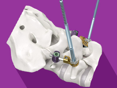
“This procedure is ideal for patients with knee pain and decreased joint function caused by small cracks or stress fractures,” explained Dr. Fish. “We inject a calcium phosphate compound that seals the crack, similar to caulking.”
Specifically, surgeons use an injectable, bone graft substitute material called AccuFill® that is intended to fill voids or gaps in the skeletal system. It flows readily to fill bone defects, then crystallizes and sets to form a scaffold in the bone. The compound promotes new bone growth and is naturally replaced with the new bone during the healing process. The FDA approved the material in 2010.
According to the Centers for Disease Control and Prevention (CDC), 719,000 Americans had a full knee replacement in 2010. That same year, the National Center for Health Statistics released a report that shows hospitalization rates for knee replacements more than doubled over a ten year period for adults aged 55-64.
Dr. Fish and his partner Dr. Robert Daher, and colleague Dr. Matthew Rogell, have offered the procedure for the past two years.

“Before Subchondroplasty, there were limited treatment options for this type of injury,” said Dr. Rogell. “Often the only treatment option for patients in significant pain was a more invasive knee replacement.”
For Saccone, his operation was performed at 7:00 a.m. and by noon he was back in his home and able to move around.
“On just the fourth day after my surgery, I was walking without a cane. I couldn’t believe it.” Saccone said. Eight months later, Saccone remains very active, walking 1 ½ miles per day. He just returned from a trip to Atlantic City where he easily walked up and down the boardwalk.
The minimally invasive surgery is a 30-minute outpatient procedure performed under regional anesthesia, so the patient remains awake. The knee can be quite uncomfortable for 24-48 hours after the procedure, but then the pain resolves rapidly. Patients are on crutches for three to five days postoperatively. Most see a complete recovery in four to six weeks.

“Studies have indicated that the benefits can last for more than five years,” Dr. Fish said. “Additionally, this procedure will not limit treatment options for a partial or full joint replacement further down the road if needed.”
Dr. Fish is an examiner for the American Board of Orthopedic Surgery as well as an editorial reviewer for The Journal of Bone & Joint Surgery and the American Journal of Sports Medicine.

“Reviewing the latest research for publication in national orthopedic journals,” he said, “allows me to stay current while evaluating the risks and benefits of new technologies.” Dr. Fish has not yet performed the Subchondroplasty procedure on other weight-bearing joints such as the hip or ankle but he said the potential exists.
Dr. Fish and Dr. Daher are partners at Orthopedic Specialists of Connecticut, based in Brookfield, CT. They both reside in Ridgefield, CT. Learn more by visiting www.ctorthopaedic.com. Dr. Rogell is a partner with Connecticut Family Orthopedics based in Danbury. He resides in Redding, CT. For more information about Dr. Rogell, contact www.cfortho.org.

Western Connecticut Health Network (WCHN) is the region’s premier, patient-centred health care organization built for the people we serve in Western Connecticut and adjacent New York. WCHN is anchored by three nationally recognized hospitals, Danbury Hospital, New Milford Hospital and Norwalk Hospital, with the continuum of outpatient health and wellness services offered by numerous medical practices and sub-specialties across the region through the Western Connecticut Medical Group, the Western Connecticut Home Care. Committed to learning and innovation, our hospitals collaborate with the University of Vermont Medical College and many other well-known academic institutions to promote the most progressive care possible. The nationally renowned WCHN Research Institute, the WCHN Foundation and Norwalk Hospital Foundation and other affiliates complete the WCHN family where We Know You Well! For more information, visit TheNewWCHN.org. Share your comments with us at Facebook.com/DanburyHospital; Facebook.com/NewMilfordHospital and/or Facebook.com/NorwalkHospital.


anat_276_02_FNL.jpg

September 27, 2016 OrthoSpineNews

(DANBURY, CT ) – September 9, 2016 – Every year, 31 million Americans grumble “Oh my aching back”. Low back pain can range from a dull, constant ache to a sudden, sharp sensation that leaves the person incapacitated. Pinpointing the source of the pain and proper treatment is complicated enough, but what if the pain isn’t really caused by a problem with the spine?
Dr. David A. Bomback, a Spine and Scoliosis Surgeon with Connecticut Neck and Back Specialists, LLC in Danbury and performs surgery at Danbury Hospital, is helping patients find relief by treating a little known condition called sacroiliac joint dysfunction. Dr. Bomback is the first surgeon at Danbury Hospital to treat the condition with a sacroiliac joint fusion procedure and one of only a few surgeons in the entire state.

Where is the sacroiliac joint?
The sacroiliac joint connects the sacrum, the lowest bony segment of the spine, with the pelvis. The sacroiliac joint transmits most of the load from the upper body to the lower limbs, acting as a shock absorber in relieving forces on the spine. The joint is reinforced and supported by very strong ligaments. Occasionally, these ligaments can become too loose or too stiff so that the joint itself becomes arthritic, leading to persistent pain.
What causes sacroiliac joint dysfunction?
Sacroiliac issues are most commonly seen in mothers who have ligament loosening due to pregnancy, patients who have suffered trauma from an injury or accident, patients with certain arthritic conditions, and patients with previous lower back surgery (especially previous lumbar fusions). Approximately 25% of patients who visit spine surgeons actually have sacroiliac pain as that cause of his or her back pain rather than true lumbar (lower back pain).


Tough to Diagnose
Unfortunately, most tests including MRIs, x-rays, and CAT scans do not detect sacroiliac joint dysfunction, making the diagnosis challenging. “The diagnosis is truly made by sitting down and talking to the patient and getting an appropriate history, as well as a physical exam consistent with sacroiliac joint dysfunction,” explains Dr. Bomback. “When I examine a patient with sacroiliac pain, I stress the sacroiliac joint in a number of ways to see if I can elicit and/or reproduce the patient’s pain.”
Common Causes of Back Pain
The vast majority of low back pain is mechanical in nature, meaning damage to a vertebra of the lower spine. In many cases, it is caused by general degeneration of the spine associated with normal wear and tear that occurs in the joints, discs, and bones of the spine as people get older. Sprains, strains and traumatic injury are also contributors.
According to the National Institute of Neurological Disorders and Stroke, about 80% of adults experience low back pain at some point in their lifetimes. It is the most common cause of job-related disability and a leading contributor to missed work days. About 20% of back pain sufferers develop chronic pain with persistent symptoms lasting over one year. In some cases, treatment successfully relieves chronic pain, but in other cases pain persists despite medical and surgical treatment.
“Spinal surgeries at times can have a poor outcome if the diagnosis is made prematurely or incorrectly,” notes Dr. Bomback. “It is critical that the diagnosis of sacroiliac dysfunction is made appropriately, often times after diagnostic injections.”

The nuts and bolts of sacroiliac joint fusion surgery

The surgery involves a minimally invasive approach, placing three screws across the sacroiliac joint, bridging the spine to the pelvis to eliminate any abnormal motion thus allowing the joint to fuse over time. The surgery significantly reduces and oftentimes completely relieves the pain. An incision less than 1 inch in size is made through a tiny tube guided by a live x-ray. The surgery takes approximately 30 minutes and there is minimal and often no blood loss. The patient can usually go home the same day or occasionally after a one night hospital stay.

“The procedure has been around for approximately five to seven years,” said Dr. Bomback. “To date, approximately 19,000 patients have had the surgery in this country. We feel it is likely a permanent solution to the cause of their lower back pain and therefore offers an improvement in their quality of life.”

For more information on sacroiliac joint fusion surgery or to learn if you are a candidate for the procedure, please call 203-744-9700 or visit www.ctneckandback.com.

About Western Connecticut Health Network
Western Connecticut Health Network (WCHN) is the region’s premier, patient-centred health care organization built for the people we serve in Western Connecticut and adjacent New York. WCHN is anchored by three nationally recognized hospitals, Danbury Hospital, New Milford Hospital and Norwalk, with the continuum of outpatient health and wellness services offered by numerous medical practices and sub-specialties across the region through the Western Connecticut Medical Group, the Western Connecticut Home Care. Committed to learning and innovation, our hospitals collaborate with the University of Vermont Medical College and many other well-known academic institutions to promote the most progressive care possible. The nationally renowned WCHN Research Institute, the WCHN Foundation and Norwalk Hospital Foundation and other affiliates complete the WCHN family where We Know You Well! For more information, visit TheNewWCHN.org. Share your comments with us at Facebook.com/DanburyHospital; Facebook.com/NewMilfordHospital and/orFacebook.com/NorwalkHospital


Device_MainstayMedicalReActiv8_WEB.jpg

September 27, 2016 OrthoSpineNews

27th September 2016

Mainstay Medical has announced the one-year results from the ReActiv8-A Clinical Trial, an international, multi-centre, prospective, single arm trial for ReActiv8 in people with disabling chronic low back pain and few other treatment options.

The one-year results show sustained performance in the ReActiv8-A Clinical Trial at the one-year follow-up with a clinically important, statistically significant and lasting improvement in the study’s key endpoints for pain (NRS), disability (ODI) and quality of life (EQ-5D).

Peter Crosby, chief executive officer of Mainstay, says, “We are very encouraged to see such strong and lasting benefits in this difficult-to-treat population. After one year of ReActiv8 treatment, 88% of subjects reported a clinically important improvement in one or more of the study endpoints, 81% were satisfied or very satisfied with the treatment and the majority continued to use the ReActiv8 treatment.”

The results presented are based on data from the first 47 subjects implanted in the ReActiv8-A Trial of whom 46 have completed the 90-day follow-up, 45 the 180-day follow-up and 41 the one-year follow-up.

To facilitate future comparison of results in the ReActiv8-A and the ReActiv8-B trial, all outcomes are presented relative to the data collected at the enrolment visit, according to a company release.

Results for all subjects at 90 days, 180 days and 1 year respectively are:

  • Ninety-three per cent, 87% and 88% with clinically important improvement in one or more of the study’s key endpoints.
  • Sixty-three per cent, 58% and 56% with clinically important improvement in low back pain NRS on the day.
  • Fifty per cent, 53% and 59% with clinically important improvement in ODI.
  • Eighty-nine per cent, 82% and 80% with clinically important improvement in EQ-5D.
  • Sixty-one per cent, 67% and 62% reported>50% pain relief.
  • Eighty-nine per cent, 84% and 81% were satisfied with ReActiv8 treatment.

The results for EQ-5D and ODI previously announced were relative to data collected at the pre-implant visit to 90 days and 180 days and were:

  • Fifty-sevent per cent and 60% with clinically important improvement in ODI.
  • Sixty-seven per cent and 73% with clinically important improvement in EQ-5D.

Adverse events incidence and type were comparable to those in clinical trials reported for other neurostimulation devices, according to the release, with no unanticipated adverse events, and no serious adverse events related to the device, therapy or procedure.


bandeau_s12016_page_va.png

September 27, 2016 OrthoSpineNews

September 27, 2016 – ERAGNY-SUR-OISE, France–(BUSINESS WIRE)

SAFE ORTHOPAEDICS (Paris:SAFOR) (FR0012452746 – SAFOR), a company offering innovative ranges of sterile implants combined with their single-use instruments has announced its half-year 2016 results for the six months to June 30, 2016, as approved by the Board of Directors on September 27, 2016.

Pierre Dumouchel, Chief Executive Officer of Safe Orthopaedics, said: “In parallel with the implementation of our new strategy of refocusing on regions growing most rapidly, we introduced a cost-cutting policy in the first six months of 2016, which will deliver greater benefits starting in the second half of 2016. We are also continuing our international expansion drive. We moved into new territories and are working hard to raise the visibility of our products in the scientific community, so that the use of single-use instruments for back surgeries gains further traction.”

in thousands of euros H1 2016 H1 2015
Adjusted revenue* 1,204 1,045
Revenue 1,303 1,307
Purchases used and change in inventories (981) (894)
External costs (1,362) (1,141)
Personnel costs (2,010) (1,983)
Other operating expenses (351) (308)
Operating income/(loss) before non-recurring items (3,402) (3,019)
Operating income/(loss) (3,412) (3,019)
Net financial income/(expense) (158) 301
Net income/(loss) (3,559) (2,727)

Adjusted first-half 2016 revenue up 15%

Following the introduction of the new strategy unveiled by Safe Orthopaedics in March of refocusing on its fastest-growing regions, it recorded adjusted revenue of €1.2 million in the first half of 2016. This represented an increase of 15%, with balanced contributions from France and the Rest of the World. Including the contribution of €0.26 million from the United States in the first half of 2015, total revenue was stable at €1.3 million compared with the year-earlier period.

Safe Orthopaedics has consolidated its sales performance in France and achieved major progress in the Rest of the World.

In parallel, Safe Orthopaedics continued to expand its international distribution network by entering into additional agreements covering Australia, New Zealand and two initial countries in Latin America (Chile and Mexico). Further territories are also expected to be added during the second half of the year.

Launch of a drive to cut operating costs

Safe Orthopaedics’ first-half 2016 operating loss came to €3.4 million, compared with a loss of €3.0 million in the same period of 2015. This increase included €0.4 million in non-recurring expenses incurred as a result of refocusing in France and the Rest of the World and the departure in June of the previous Chief Executive Officer.

After taking into account €0.16 million in net financial expense as a result of currency effects on intragroup cash balances, Safe Orthopaedics recorded a first-half 2016 net loss of €3.6 million, compared with a loss of €2.7 million in the first six months of 2015.

During the period, Safe Orthopaedics launched a cost-cutting policy, which should start to pay off from the second half of 2016.

Stronger cash position

At June 30, 2016, Safe Orthopaedics held €4.3 million in cash. This does not include a €0.5 million subscription by IdInvest Partners in early July following the Shareholders’ Meeting on June 30, 2016 and another €0.5 million subscription raised from IdInvest Partners in the form of OCABSA bonds on July 26, 2016.

Next Press Release: Q3 2016 revenue, October 13, 2016 (after market close)

About Safe Orthopaedics

Founded in 2010, Safe Orthopaedics is a French medical technology company that develops and markets an innovative range of sterile implants and associated single-use surgical instruments, with the aim of facilitating safer, optimized and lower-cost spinal surgery. By avoiding the reuse of surgical instruments, Safe Orthopaedics reduces the risk of infection, avoids the cumbersome and unreliable logistics of instrument sterilization, and limits hospital costs. Protected by 17 patent families, the SteriSpine™ Kits are CE-marked and FDA cleared. The company is based at Eragny-sur-Oise (France), and has 34 employees. For more information, visit:www.SafeOrtho.com

* Adjusted for operations in the United States discontinued effective March 1, 2016.

Contacts

Safe Orthopaedics
Thierry Lambert, +33 (0)1 34 21 50 00
CFO
investors@safeorthopaedics.com
or
NewCap
Julien Perez / Valentine Brouchot
Investor Relations
or
Nicolas Merigeau
Media Relations
Tel. : +33 (0)1 44 71 94 94
SafeOrtho@newcap.eu


Syzygy_image1.jpg

September 27, 2016 OrthoSpineNews

MARIETTA, GA (PRWEB) SEPTEMBER 27, 2016

Balanced spinal solutions provider Amendia Inc. is pleased to announce the commercial launch of its innovative Spondylolisthesis reduction system:, the Syzygy™ Stabilization System. Configured to provide immobilization and stabilization of spinal segments as an adjunct to fusion, Syzygy is ideal for use in the treatment of specific acute and chronic instabilities or deformities of the thoracic, lumbar, or sacral spine; and conditions such as fracture, dislocation, scoliosis, kyphosis, spinal tumor, or failed previous fusion.

Made of implant-grade titanium alloy, the Syzygy system features cannulated and non-cannulated screws, with self-tapping “grip quick” threads for maximum cortical bone purchase. Extended threads allow increased adjustability and control in 5mm increments for accurate reduction of spondylolisthesis. With available medial and cranial couplers, Syzygy facilitates up to 35mm of reduction with ±10° of cranial-caudal adjustment.

Dr. Hewatt M. Sims, MD, fellowship-trained orthopedic spine surgeon at Orthopedic Spine Center and Tift Regional Medical Center in Tifton, GA, performed recent spine surgeries with Syzygy. “Syzygy is a unique spinal system, providing quick, precise, and consistent reduction of the spondylolisthesis deformity,” Dr. Sims said.

The Syzygy system’s advanced design features, such as extended threads on each MAC Pin enable precise corrections and dual scales enhance optimal sagittal alignment.

“In working hand in hand with the medical community and spinal device marketplace, we know how important it is to provide patients and surgeons with a pedicle screw-based solution that consistently and accurately realigns the spine,” said Lawrence Boyd, Amendia’s Executive Vice President of Research & Development. “With surgeons providing care for a large number of patients who engage in athletics and rigorous physical activity, combined with a growing aging population, this critical addition to our line of lumbar fixation products will provide another way for surgeons to evolve and innovate their care.”

The Syzygy Stabilization system is part of Amendia’s growing list of products for balanced solutions for lumbar fixation systems. To learn more about Syzygy, including features and benefits and 510(k) information, please visit us at http://www.amendia.com.

About Amendia

Headquartered in a state-of-the-art manufacturing facility in Marietta, GA, Amendia is a leading designer, developer, manufacturer and marketer of medical devices used in spinal surgical procedures. Amendia’s mission is to exceed surgeon and patient expectations by creating balanced solutions with disruptive technologies for medical devices paired with biologics and instrumentation. Amendia’s vertically-integrated strategy focuses on improving surgical outcomes and the lives of patients with spinal disorders. For more information, please visit http://www.amendia.com.


LineUP-combined.png

September 27, 2016 OrthoSpineNews

A new orthopedic CT system promises to improve the standard of care for knee imaging. On the CurveBeam LineUp, patients are scanned while standing upright and fully weight bearing.

Traditional CT and MR images are acquired in a non-weight bearing position, leading to “missed diagnoses of meniscal damage,” according to Dr. Neil Segal, who has been overseeing research efforts using a LineUp prototype, first at the University of Iowa and currently at the University of Kansas.

Although plain radiographs can be acquired while the patient is in a full weight-bearing position, the optimal degree of knee flexion and X-Ray beam tilt to best visualize the joint surface is person specific.

“Difficulty in reproducing the same view of the joint over time impairs ability to detect joint disease, and the 2D nature of radiographs makes these images of overlapping bony anatomy very insensitive for detecting abnormalities until there is advanced joint damage,” Dr. Segal said.

The LineUp was developed by CurveBeam, a Pennsylvania-based company that specializes in extremity cone beam CT systems for orthopedics. CurveBeam anticipates it will submit an application for and receive FDA 510(k) clearance for the LineUp in 2017. The LineUp will be on display at RSNA at Booth #8008 in the North Hall.

CurveBeam introduced the pedCAT, a bilateral weight bearing CT system dedicated to the foot and ankle, in 2012. Since then, the device has been added to the imaging services of hospital foot & ankle sections, orthopedic clinics and podiatry offices.

Like the pedCAT, the LineUp will provide isotropic, three-dimensional volumes of the anatomy with a high resolution output of between 0.2 mm and 0.3 mm slices. The LineUp will be the only cone beam CT system that can provide bilateral, weight bearing scans.

A study led by Dr. Segal focused on osteophytes, one structure linked to pain in people with knee osteoarthritis. Knees of community-dwelling adults with knee OA were imaged with MRI (reference), fixed-flexion radiographs, and weight bearing CT. The sensitivity and accuracy for detecting osteophytes and subchondral cysts were higher with weight bearing CT imaging in comparison to fixed-flexion radiographs. The study was published in the August 2016 issue of the Journal of Orthopedic Research.

“Clinically, this is a highly meaningful improvement,” Dr. Segal said. “It suggests that weight-bearing CT could replace radiographs as the recommended means of assessing knee OA. This advancement is even more significant given that it was made without significantly increasing the radiation dose (0.01 mSv for SCT vs. 0.005–0.102 mSv for a series of knee radiographs).”

Another research effort led by Dr. Segal indicates weight-bearing CT arthrography studies can provide outstanding delineation of articular cartilage with better differentiation between cartilage and subchondral bone then MRI studies, while also visualizing the cruciate ligaments. In knees with osteoarthritis, meniscal tears not visualized on MRI were detectable on weight-bearing CT.

“Thus, we found that some potential advantages of weight-bearing CT over non-weight-bearing MRI/MRA include 3D measures of meniscal position, detection of pathology not detected in unloaded positions, and ability to bear weight in a functional position, thus better recreating the magnitude of body weight and muscle forces acting around the knee during usual standing,” Dr. Segal said.


Spineology_Logo_2014-1-1200x332.jpg

September 27, 2016 OrthoSpineNews

September 27, 2016

MINNEAPOLIS & ST. PAUL, Minn.–(BUSINESS WIRE)–Spineology Inc., the innovator in anatomy-conserving spine surgery, announced today the availability of multiple transforaminal lumbar interbody fusion (TLIF) technologies as part of the Anatomy-Conserving Medial Lumbar Interbody Fusion (ACT MediaLIF™) platform. These TLIF technologies allow for TLIF placement of interbody devices using a direct posterior, or ACT MediaLIF, approach.

“Surgeons frequently prefer placing a single cage using the TLIF technique over more traditional PLIF techniques, but experience difficulty with TLIF when using the latest mini-midline techniques,” said John Booth, CEO of Spineology Inc. “These unique technologies allow surgeons to use a TLIF technique and still take advantage of the benefits that the ACT MediaLIF procedure provides.”

Dr. Vivek Mohan, MD, of Dupage Medical Group in Chicago added, “Having technology that allows me to do a TLIF in conjunction with the ACT MediaLIFprocedure provides the best solution for my patients. The procedure allows me to reduce the incision size and muscle trauma, and minimize the amount of bony removal to place the cage. It also facilitates anterior cage placement to maximize lordosis restoration.”

The ACT MediaLIF procedure combines Spineology’s Medius Retractor, Rampart Interbody and Fortress Pedicular Fixation Systems to provide an anatomy-conserving approach to posterior lumbar surgery that can reduce incision length, dissection and retraction requirements compared to traditional open surgery.

About Spineology Inc.
Spineology Inc. provides innovative, anatomy-conserving spinal technologies for surgeons and their patients. Spineology surgical techniques conserve spinal bone, ligament and muscle tissue. Spineology is committed to increasing procedural efficiency, reducing surgical morbidity and accelerating patient recovery. Learn more at spineology.com.

Contacts

Spineology Inc.
John Booth, 651-256-8511
jbooth@spineology.com
or
Risdall Public Relations
Dave Folkens, 651-286-6713
dave@risdall.com


joimax_Logo_highres.jpg

September 26, 2016 OrthoSpineNews

September 26, 2016

IRVINE, Calif.–(BUSINESS WIRE)–The AMA (American Medical Association) released the CPT 2017 Codebook (Current Procedural Terminology), now containing the first endoscopic spinal surgery code no. 62380. This new code covers endoscopic decompression of the spinal cord, nerve root(s), including laminotomy, partial facetectomy, foraminotomy, discectomy and/or excision of herniated intervertebral disc, 1 interspace, lumbar, and will be enforced as of January 1st, 2017. Under this code the specific transforaminal (TESSYS®) and/or interlaminar (iLESSYS®) procedures are fully covered in the USA herewith, which are already marketed by joimax® in 45 countries.

“The definition of this new code in the CPT codebook 2017 is a major milestone towards the acceptance of endoscopic minimally invasive spine surgery techniques and its benefits to both the surgeon and their patients,” states Wolfgang Ries, Founder and CEO of joimax®. Since 2012 joimax® had been driving this effort to obtain CPT codes as these procedures required submission to payors for reimbursement coverage which was granted only in single cases under temporary codes, so-called T-codes.

“joimax® has been instrumental in driving the effort to obtain full code coverage of endoscopic spinal surgery,” says Daniel Laich, DO, Swedish Convenant Hospital, Chicago, who had been participating and presenting in almost every AMA meeting over the last five years. Due to the high research and publication activities by key users like Albert Telfeian, MD, PhD, at Rhode Island Hospital at Brown University, and a group of users around him, strong evidence has been accepted by AMA authorities, justifying the release of this new CPT code.

Many papers and publications on joimax® products can now be downloaded from the newly released joimax® homepage. The completely redesigned website offers visitors richer insight into the company’s innovative products and systems. A quick and easy access to essential information and features is available, including the latest workshop and education activities, which are essential in endoscopic minimally-invasive spinal surgery.

Meanwhile joimax® offers basic and advanced workshop training courses mainly in the USA, Germany and Austria, and oversees a participation population of almost 3,000 physicians. Independent training courses are also performed in China and Korea to support the fast growing Asian markets. Detailed information about those courses are updated regularly at www.joimax.com.

About joimax®

Founded in Karlsruhe, Germany, in 2001, joimax® is the leading developer and marketer of complete systems for endoscopic minimally invasive spinal surgery. With TESSYS® (transforaminal), iLESSYS® (interlaminar) and CESSYS® (cervical) for decompression procedures, Multiuse RT (e.g. for rhizotomy) or with EndoLIF® and Percusys® for minimally-invasive endoscopic assisted stabilizations, proven endoscopic systems are provided that, together, cover a variety of indications.

In procedures for herniated disc, stenosis, pain therapy or spinal stabilization treatment, surgeons utilize joimax® technologies to operate through small incisions – under local or full anesthetic – via tissue and muscle-sparing corridors through natural openings into the spinal canal (e.g. intervertebral foramen, the “Kambin triangle”).

Contacts

joimax® GmbH
Sabine Jarosch, 49 721 25514 0
sabine.jarosch@joimax.com


8-SonicOne-OR-small-1200x760.png

September 26, 2016 OrthoSpineNews

FARMINGDALE, N.Y., Sept. 26, 2016 /PRNewswire/ — Misonix, Inc. (NASDAQ: MSON), an international ultrasonic surgical device company that designs, manufactures and markets innovative therapeutic ultrasonic instruments for spine surgery, neurosurgery and other surgical specialties, today announced new data demonstrating the effectiveness of the SonicOne O.R. ultrasonic debridement device in removing biofilm from potentially contaminated surfaces associated with metal knee and hip implants. The data will be presented during the World Union of Wound Healing Societies (‘WUWHS”) to be held September 25 – 29, 2016 in Florence, Italy.

It is estimated that 90% of chronic wounds contain bacteria and fungi living within biofilm. The new data suggests SonicOne O.R. can play a role in addressing the significant problem biofilm plays in preventing chronic wounds from healing.

“The early data are very encouraging. The studies showed complete removal of biofilm from infected titanium plates. The plates simulated biofilm that grows on metal knee and hip implants. Biofilm formation and removal is a significant problem in infected implants,” said Dr. Mark S. Granick, Professor and Chief, Division of Plastic Surgery, Rutgers New Jersey Medical School. Dr. Granick added, “While more studies are needed, SonicOne continues to demonstrate utility as an infection control platform during wound debridement.”

Stavros Vizirgianakis, Interim Chief Executive Officer of Misonix, commented, “We are pleased that the SonicOne O.R. system is being recognized as a viable solution to a recurring problem that can profoundly impact the quality of life for the growing number of implant recipients throughout the world. It is gratifying that globally recognized experts like Dr. Granick are involved in this ongoing research. The SonicOne O.R. features unique attributes that can enhance healing in diabetic ulcers, wounds from burns and now, potentially, implant infections. We look forward to the forthcoming results of the ongoing research.”

Misonix also announced a Live YouTube event from Florence, Italy featuring Dr. Granick’s presentation during the World Union Wound Healing Society Meeting. The Live event can be viewed at https://www.youtube.com/embed/VZ3udLGk0e4 on Monday, September 26th at 5 pm EST.  The presentation is expected to cover the clinical benefits of the use of the newly launched SonicVac® addition to the SonicOne O.R. device.  The SonicVac, which was launched in April of this year, uniquely combines aspiration and ultrasound during wound debridement. This combination reduces spray during the procedure while capturing debris and bacteria from the wound.

“We’re particularly excited that Dr. Granick is hosting this event and we look forward to his live presentation at WUWHS 2016 for a worldwide audience.  Events like this are an important part of the Misonix education platform and will expose clinicians from around the world to our unique technologies,” states Scott Ludecker, Senior Vice President, Global Sales and Marketing with Misonix.

The SonicOne O.R. System is an innovative ultrasonic surgical debridement system that allows surgeons to address the challenges chronic wounds present to them, the patient and the healthcare system. SonicOne O.R. establishes a new standard in surgical wound bed preparation, an essential first step in the wound healing process.  The new SonicOne procedure will be sold and distributed through the Misonix sales team in the United States and its domestic and international distribution networks.

About Misonix
Misonix, Inc. designs, develops, manufactures and markets therapeutic ultrasonic medical devices. Misonix’s therapeutic ultrasonic platform is the basis for several innovative medical technologies.

Addressing a combined market estimated to be in excess of $1.5 billion annually; Misonix’s proprietary ultrasonic medical devices are used in spine surgery, neurosurgery, orthopedic surgery, wound debridement, cosmetic surgery, laparoscopic surgery, and other surgical and medical applications.  Additional information is available on the Company’s Web site atwww.misonix.com.

Corporate Contact
Misonix Contact:  Investor Contact:
Richard Zaremba Joe Diaz
631-694-9555 Lytham Partners
invest@misonix.com 602-889-9700
info@misonix.com


Safe Harbor Statement
With the exception of historical information contained in this press release, content herein may contain “forward looking statements” that are made pursuant to the Safe Harbor Provisions of the Private Securities Litigation Reform Act of 1995. These statements are based on management’s current expectations and are subject to uncertainty and changes in circumstances. Investors are cautioned that forward-looking statements involve risks and uncertainties that could cause actual results to differ materially from the statements made. These factors include general economic conditions, delays and risks associated with the performance of contracts, risks associated with international sales and currency fluctuations, uncertainties as a result of research and development, acceptable results from clinical studies, including publication of results and patient/procedure data with varying levels of statistical relevancy, risks involved in introducing and marketing new products, potential acquisitions, consumer and industry acceptance, litigation and/or court proceedings, including the timing and monetary requirements of such activities, the timing of finding strategic partners and implementing such relationships, regulatory risks including approval of pending and/or contemplated 510(k) filings, the ability to achieve and maintain profitability in the Company’s business lines, the completion of the investigation related to identified deficiencies in internal control over financial reporting, and other factors discussed in the Company’s Annual Report on Form 10-K, subsequent Quarterly Reports on Form 10-Q and Current Reports on Form 8-K. The Company disclaims any obligation to update its forward-looking relationships.

Logo – http://photos.prnewswire.com/prnh/20160201/328020LOGO


SOURCE Misonix, Inc.

Related Links

http://www.misonix.com


stryker-2-big.jpg

September 26, 2016 OrthoSpineNews

Kalamazoo, Michigan – September 26, 2016 – Stryker Corporation (SYK) announced that it will host a conference call on Thursday, October 27, 2016 at 4:30 p.m., Eastern Time, to discuss the Company`s operating results for the quarter ended September 30, 2016 and provide an operational update. Final operating results for the quarter ended September 30, 2016 will be released at 4:00 p.m. that day.

 To participate in the conference call dial (844) 826-0610 (domestic) or (973) 453-3249 (international) and be prepared to provide conference ID number 57981183 to the operator.

A simultaneous webcast of the call will be accessible via the Company`s website at www.stryker.com. The call will be archived on the Investors page of this site.

A recording of the call will also be available from 8:00 p.m., Eastern Time, on Thursday, October 27, 2016, until 11:59 p.m., Eastern Time, on Thursday, November 3, 2016. To hear this recording you may dial (855) 859-2056 (domestic) or (404) 537-3406 (international) and enter conference ID number 57981183.

Forward-looking statements
This presentation may contain information that includes or is based on forward-looking statements within the meaning of the federal securities law that are subject to various risks and uncertainties that could cause our actual results to differ materially from those expressed or implied in such statements. Such factors include, but are not limited to: weakening of economic conditions that could adversely affect the level of demand for our products; pricing pressures generally, including cost-containment measures that could adversely affect the price of or demand for our products; changes in foreign exchange markets; legislative and regulatory actions; unanticipated issues arising in connection with clinical studies and otherwise that affect U.S. Food and Drug Administration approval of new products; changes in reimbursement levels from third-party payors; a significant increase in product liability claims; the ultimate total cost with respect to the Rejuvenate and ABG II matter; the impact of investigative and legal proceedings and compliance risks; resolution of tax audits; the impact of the federal legislation to reform the United States healthcare system; changes in financial markets; changes in the competitive environment; our ability to integrate acquisitions; and our ability to realize anticipated cost savings. Additional information concerning these and other factors is contained in our filings with the U.S. Securities and Exchange Commission, including our Annual Report on Form 10-K and Quarterly Reports on Form 10-Q.

Stryker is one of the world`s leading medical technology companies and, together with our customers, we are driven to make healthcare better. The Company offers a diverse array of innovative products and services in Orthopaedics, Medical and Surgical, and Neurotechnology and Spine that help improve patient and hospital outcomes. Stryker is active in over 100 countries around the world.  Please contact us for more information at www.stryker.com.

Contacts

For investor inquiries please contact:
Katherine A. Owen, Stryker Corporation, 269-385-2600 or katherine.owen@stryker.com

For media inquiries please contact:
Yin Becker, Stryker Corporation, 269-385-2600 or yin.becker@stryker.com

This announcement is distributed by NASDAQ OMX Corporate Solutions on behalf of NASDAQ OMX Corporate Solutions clients.

The issuer of this announcement warrants that they are solely responsible for the content, accuracy and originality of the information contained therein.
Source: Stryker Corporation via GlobeNewswire