Park City, UT

3 days / 6 sessions
Current Issues in Spine

February 2-4, 2017

1-7-1.jpg

January 29, 2018 OrthoSpineNews

NEW YORKJan. 29, 2018 /PRNewswire/ — Paradigm Spine, LLC, a leader in providing motion preservation solutions for the treatment of lumbar spinal stenosis, today announced publication of the ESCADA 2-year trial results (European Study of coflex And Decompression Alone) in the January 2018 issue of the Journal of Neurosurgery Spine (JNS), official journal of the American Association of Neurological Surgeons (AANS). ESCADA reports a prospective, randomized, controlled, multi-center trial comparing decompression alone to decompression with coflex® Interlaminar Stabilization® for treatment of moderate to severe lumbar spinal stenosis.  coflex is the first and only motion preserving minimally invasive treatment for moderate to severe spinal stenosis post decompression.  A total of 225 patients were enrolled in the study with 91% follow up at 2 years.

The study authors concluded that decompression with coflex Interlaminar Stabilization extends the durability and sustainability of a decompression procedure.

“These data strongly validate for the surgical spine community that coflex is a safe and effective protector of a decompression procedure,” said Marc Viscogliosi, President and CEO of Paradigm Spine.  “This means that surgeons treating patients with lumbar spinal stenosis can be more confident than ever in their treatment options, knowing that a randomized controlled trial has demonstrated with statistical significance that nearly twice as many patients with decompression alone have a risk of secondary intervention as compared to coflex patients. coflex is the first and only lumbar spinal device that has produced the highest level of evidence in two separate prospective, randomized, controlled studies against two different control groups – which exhibits the thoroughness and reproducibility of these findings. There is now potentially a more effective surgical option between the two typical treatments for lumbar spinal stenosis, and we are proud to lead the way in changing the standard of care for surgeons and their patients with this diagnosis.”

Highlights of ESCADA results:

  • The rate of patients achieving Composite Clinical Success was statistically superior in the coflex group vs. the Decompression Alone group (p=0.017)
  • Patients who received Decompression Alone experienced significantly more loss of foraminal and posterior disc height maintenance compared to the coflex group (p<0.001)
  • The Decompression Alone group experienced 228% more subsequent epidural steroid injections compared to the coflex group (p=0.0065)
  • coflex patients experienced 2.4 times more improvement in walking distance measurements compared to Decompression Alone patients (p=0.062)
  • At 24 months, the Decompression Alone group was 1.4 times more likely to be taking opioids compared to the coflex group

The ESCADA trial included seven sites in Europe / Germany with a total of 225 patients who were randomized to either decompression alone (DA, control group) or decompression with coflex Interlaminar Stabilization (coflex, study group). Overall trial success was based on a combined outcome measure called Composite Clinical Success (CCS),  in which all four components must be met: 1) ODI success with improvement > 15 points; 2) survivorship with no secondary surgical interventions or lumbar injections; 3) neurologic maintenance or improvement without worsening; 4) no device-related severe adverse events. This is the same success criteria that was used in the U.S. IDE study of coflex that has published 5-year follow-up, allowing some direct comparisons to be drawn between the cohorts representing 547 patients collectively.  Secondary measures were also collected such as walking distance, foraminal and disc height maintenance, leg and back pain scores, and opioid use.

“With the important work of the ESCADA trial, we can see that coflex extends the success and durability of a decompression procedure for lumbar spinal stenosis, while still allowing natural motion to occur,” says ESCADA author and Principal Investigator Professor Michael Rauschmann. “The U.S. IDE study first investigated and defined the long-term problems with lumbar fusion procedures that can be alleviated with coflex.  And now, through this controlled trial, we can begin to answer the questions of the sustainability of decompression alone, and the need to provide additional stabilization for those patients with coflex at first surgical intervention to avoid future complications.”

Journal of Neurosurgery Spine. January 2018. Volume 28, Issue 1 (http://thejns.org/doi/full/10.3171/2017.11.SPINE17643).

About Lumbar Spinal Stenosis (LSS)
Lumbar spinal stenosis (LSS), affecting 1.6 million patients annually in the United States, is a debilitating and degenerative disease often associated with significant leg and back pain, leg numbness and weakness, and significant reduction in an active lifestyle. Historically, the two traditional surgical treatment options for LSS included decompression alone or decompression with lumbar fusion.  Decompression alone has proven effective at relieving pain symptoms caused by lumbar spinal stenosis, however, patients may not experience long term symptomatic relief, resulting in subsequent epidural injections for pain management, or additional surgeries for conversion to a fusion. Decompression with fusion has proven to provide pain relief and stabilize the diseased segment, but may lead to adjacent level disease requiring subsequent surgeries.

About Paradigm Spine, LLC:
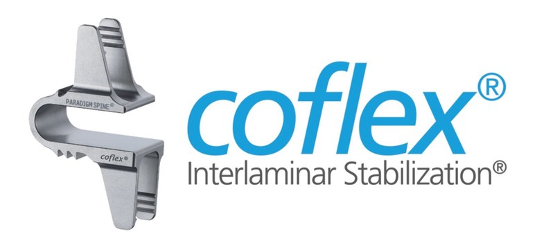
Paradigm Spine, LLC, founded in 2004, is a privately held company and remains focused on the design and development of solutions for the disease management of spinal stenosis.  The Company’s signature product is the coflex® Interlaminar Stabilization® device, which is currently used in over 50 countries worldwide. coflex is the only lumbar spinal device that has produced Level I evidence in two separate prospective, randomized, controlled studies against two different control groups, changing the standard of care for lumbar spinal stenosis treatment. For additional information visit www.paradigmspine.com or www.coflexsolution.com.


SOURCE Paradigm Spine, LLC

Related Links

http://www.paradigmspine.com


20170503_foot-and-ankle_86290.jpg

January 26, 2018 OrthoSpineNews

Further Key Findings From the Study Suggest:

  • Bracing and support dominated the market in 2016. Increasing application of braces drives the market and has resulted in industry players developing and introducing new products in the market. For instance, in October 2017, Vickers Grant Holdings, LLC launched ATHLETICA with an ankle brace as an introductory product for the brand.
  • Aging results in significant changes in the skeletal and neuromuscular systems, leading to arthritis, weak ligament, and ACL tear. Symptomatic and asymptomatic tears affect more than 50% of the geriatric population. This is one of the key drivers attributed to the accelerated growth of soft tissue orthopedic devices.
  • Geographically, North America held the largest market share in 2016 and is expected to maintain its position during the forecast period. This large share can be accounted for high adoption rate of new technology and favorable reimbursement scenario.
  • The companies are tending toward providing novel products by adopting new technology. In February 2016, Zimmer Biomet received FDA approval for its 3D printed foot-and-ankle implantUnite3D Bridge Fixation System. The product is perceived to offer greater stability and rigidity due to its integrated structure.
  • Some key industry contributors are DePuy Synthes, Zimmer Biomet, Stryker, Integra LifeSciences Corporation, Wright Medical Technology, Inc., Advanced Orthopedic Solutions, Acumed, Bioretec Ltd., and Ossur.

The global foot and ankle devices market is expected to reach USD 3.4 billion by 2025

The market is expected to observe an exponential growth during the forecast period due to the rising incidence of ankle sprains, hammertoe, bunions, osteoporosis and rheumatoid arthritis.

Ankle fractures are one of the most common types of foot injury. Nearly 170,000 people suffer from ankle fractures per year, which require surgical treatment. It is one of the nine most common fractures. This can be attributed to the relatively small surface area of contact in the ankle and the 1.5 times higher body weight on it at rest & 5.5 times higher forces in case of heavy activity. This makes the long-term viability of ankle joints crucial.

Nearly 50,000 patients are diagnosed with end-stage ankle arthritis per year, wherein the ankle cartilage is worn out, causing painful bone-on-bone contact and disability. These patients are recommended total ankle replacements as opposed to ankle fusion, as it provides higher efficacy, thus driving the market. Favorable reimbursement scenario for joint replacements is one the major contributors to its market growth. Medicare, along with some private insurers, reimburses the procedure.

Key Topics Covered:

Chapter 1 Executive Summary
1.1 Market Snapshot

Chapter 2 Research Methodology
2.1 Information Procurement
2.2 Data Analysis

Chapter 3 Foot And Ankle Devices Market Variables, Trends & Scope
3.1 Market Segmentation and Scope
3.1.1 Market Driver Analysis
3.1.1.1 Increasing expenditure on research and development (R&D) by industry players
3.1.1.2 Increasing geriatric population
3.1.1.3 Increasing prevalence of target disease
3.1.1.4 Rising incidence rate of road accidents
3.1.2 Market Restraint Analysis
3.1.2.1 Uncertainty in regulatory reforms
3.1.2.2 High cost of devices
3.2 Penetration & Growth Prospects Mapping
3.3 Foot and Ankle Devices Market – PESTLE Analysis
3.4 Industry Analysis – Porter’s

Chapter 4 Foot And Ankle Devices :Product Estimates & Trend Analysis
4.1 Foot And Ankle Devices Market: Product Movement Analysis
4.2 Bracing and Support
4.3 Joint Implants
4.4 Soft Tissue Orthopedic Devices
4.5 Orthopedic Fixation
4.6 Prosthetics

Chapter 5 Foot And Ankle Devices: Application Estimates & Trend Analysis
5.1 Foot and Ankle Devices Market: Application Movement Analysis
5.2 Hammertoe
5.3 Trauma
5.4 Osteoarthritis
5.5 Rheumatoid Arthritis
5.6 Neurological Disorders
5.7 Bunions
5.8 Osteoporosis

Chapter 6 Foot And Ankle Devices: Regional Estimates & Trend Analysis, by Product and Application

Chapter 7 Competitive Landscape

For more information about this report visit https://www.researchandmarkets.com/research/gpnt3k/3_4_bn_foot_and?w=5

Media Contact:

Research and Markets
Laura Wood, Senior Manager
press@researchandmarkets.com

For E.S.T Office Hours Call +1-917-300-0470
For U.S./CAN Toll Free Call +1-800-526-8630
For GMT Office Hours Call +353-1-416-8900

U.S. Fax: 646-607-1907
Fax (outside U.S.): +353-1-481-1716

SOURCE Research and Markets

Related Links

http://www.researchandmarkets.com


invivo-therapeutics-7x4-1.jpg

January 26, 2018 OrthoSpineNews

January 26, 2018

CAMBRIDGE, Mass.–(BUSINESS WIRE)–InVivo Therapeutics Holdings Corp. (Nasdaq: NVIV) today announced that it has entered into a common stock purchase agreement with Lincoln Park Capital Fund, LLC (“Lincoln Park”), a Chicago-based institutional investor, under which the Company has the right to sell up to $15 million in shares of common stock to Lincoln Park over a twenty-four-month period, subject to certain limitations and conditions set forth in the purchase agreement and registration rights agreement, including:

  • the company, in its sole discretion, controls the timing and amount of any sales of common stock;
  • the purchase price is based on prevailing market prices with no warrants;
  • Lincoln Park cannot require the company to make sales, but is obligated to make purchases as the company directs in accordance with the terms of the purchase agreement;
  • there are no upper limits on the price per share that Lincoln Park could be obligated to pay for shares of common stock under the purchase agreement;
  • there are no financial covenants, rights of first refusal, participation rights or liquidated damages; and
  • the SEC declares effective a registration statement, registering the shares of common stock that Lincoln Park purchases pursuant to the purchase agreement.

“We are pleased to enter into this purchase agreement with Lincoln Park, which offers us financial flexibility on favorable terms to the company and its shareholders,” said Richard Toselli, M.D., Acting Chief Executive Officer. “We welcome their investment as we pursue our near-term strategic objectives and continue our discussions with the FDA regarding a randomized controlled study in acute complete thoracic spinal cord injury that evaluates the Neuro-Spinal Scaffold™ compared to standard of care.”

In consideration for entering into the purchase agreement, the company has issued shares of common stock to Lincoln Park as a commitment fee. The purchase agreement may be terminated by the company at any time, in its sole discretion.

A more detailed description of the purchase agreement and registration rights agreement is set forth in the company’s Current Report on Form 8-K as filed with the SEC which the company encourages be reviewed carefully.

About InVivo Therapeutics

InVivo Therapeutics Holdings Corp. is a research and clinical-stage biomaterials and biotechnology company with a focus on treatment of spinal cord injuries. The company was founded in 2005 with proprietary technology co-invented by Robert Langer, Sc.D., Professor at Massachusetts Institute of Technology, and Joseph P. Vacanti, M.D., who then was at Boston Children’s Hospital and who now is affiliated with Massachusetts General Hospital. In 2011, the company earned the David S. Apple Award from the American Spinal Injury Association for its outstanding contribution to spinal cord injury medicine. In 2015, the company’s investigational Neuro-Spinal Scaffold™ implant received the 2015 Becker’s Healthcare Spine Device Award. The publicly traded company is headquartered in Cambridge, MA. For more details, visit www.invivotherapeutics.com.

Safe Harbor Statement

Any statements contained in this press release that do not describe historical facts may constitute forward-looking statements within the meaning of the federal securities laws. These statements can be identified by words such as “believe,” “anticipate,” “intend,” “estimate,” “will,” “may,” “should,” “expect,” “designed to,” “potentially,” and similar expressions, and include statements regarding the status of the company’s clinical program. Any forward-looking statements contained herein are based on current expectations, and are subject to a number of risks and uncertainties. Factors that could cause actual future results to differ materially from current expectations include, but are not limited to, risks and uncertainties relating to the company’s discussions and engagement with the FDA; the company’s ability to initiate, conduct and complete clinical trials; the expected benefits and potential efficacy of the company’s products and technology in connection with the treatment of spinal cord injuries; the availability of substantial additional funding for the company to continue its operations and to conduct research and development, clinical trials and future product commercialization; and other risks associated with the company’s business, research, product development, attainment of regulatory approval, marketing and distribution plans and strategies identified and described in more detail in the company’s Quarterly Report on Form 10-K for the three months ended September 30, 2017, and its other filings with the SEC, including the company’s most recent Form 10-K, its Form 10-Qs and its current reports on Form 8-K. The company does not undertake to update these forward-looking statements.

Contacts

InVivo Therapeutics Holdings Corp
Heather Hamel, 617-863-5530
Investor Relations
Investor-relations@invivotherapeutics.com


180124111239_1_540x360-y-1-1.jpg

January 26, 2018 OrthoSpineNews

January 24, 2018-Credit: University of Sydney

A discovery by University of Sydney researchers could underpin a new class of implantable devices that provide biological signals to surrounding tissue for better integration with the body and reduced risk of infection.

Modern medicine increasingly relies on implantable biomedical devices but their effectiveness is often limited because of unsuccessful integration with host tissue or the development of untreatable infections, necessitating replacement of the device through revision surgery.

The team at the Applied Plasma Physics and Surface Engineering Laboratory has developed practical techniques to guide and attach peptides to surfaces; computer simulations and experiments demonstrated control of both peptide orientation and surface concentration, which can be achieved by applying an electric field like that delivered by a small household-sized battery.

The findings are published today in Nature Communications.

Corresponding author Professor of Applied Physics and Surface Engineering Marcela Bilek said biomaterial coatings can mask the implanted devices and mimic surrounding tissue.

“The holy grail is a surface that interacts seamlessly and naturally with host tissue through biomolecular signalling,” said Professor Bilek, who is a member of the University of Sydney Nano Institute and the Charles Perkins Centre.

Robust attachment of biological molecules to the bio-device surface is required to achieve this, as enabled by unique surface modification processes developed by Professor Bilek.

“Although proteins have successfully been used in a number of applications, they don’t always survive harsh sterilisation treatments — and introduce the risk of pathogen transfer due to their production in micro-organisms,” Professor Bilek said.

Professor Bilek — together with Dr Behnam Akhavan from the School of Aerospace, Mechanical and Mechatronic Engineering and the School of Physics and lead author PhD candidate, Lewis Martin from the School of Physics — are exploring the use of short protein segments called peptides that, when strategically designed, can recapitulate the function of the protein.

Mr Martin said the team was able to tune the orientation of extremely small biomolecules (less than 10 nanometres in size) on the surface. “We used specialised equipment to perform the experiments, but the electric fields could be applied by anyone using a home electronics kit,” he said.

Dr Akhavan said that assuming industry support and funding for clinical trials, improved implants could be available to patients within five years.

“The application of our approach ranges from bone-implants to cardiovascular stents and artificial blood vessels,” Dr Akhavan said.

“For the bone implantable devices, for example, such modern bio-compatible surfaces will directly benefit patients suffering from bone fracture, osteoporosis, and bone cancer.”

Because of their small size, the peptides can be produced synthetically and they are resilient during sterilisation. The main difficulty in using peptides is ensuring they are attached at appropriate densities and in orientations that effectively expose their active sites.

Using applied electric fields and buffer chemistry, the researchers discovered several new levers that control peptide attachment. Charge separation on peptides creates permanent dipole moments that can be aligned with an electric field to provide optimal orientation of the molecules and the amount of peptide immobilised can also be tuned by the electrostatic interactions when the peptides have an overall charge.

The paper said this knowledge is being used to design strategies to create a new generation of synthetic biomolecules.

“Our findings shed light on mechanisms of biomolecule immobilisation that are extremely important for the design of synthetic peptides and biofunctionalisation of advanced implantable materials,” the paper states.

Story Source:

Materials provided by University of Sydney

Journal Reference:

  1. Lewis J. Martin, Behnam Akhavan, Marcela M. M. Bilek. Electric fields control the orientation of peptides irreversibly immobilized on radical-functionalized surfacesNature Communications, 2018; 9 (1) DOI: 10.1038/s41467-017-02545-6

University of Sydney. “Implantable medical devices bolstered by next-gen surface modification: Synthetic peptides could integrate seamlessly with host tissue.” ScienceDaily. ScienceDaily, 24 January 2018. <www.sciencedaily.com/releases/2018/01/180124111239.htm>.


ArthroS_Movie_Picture_VirtaMed_web555-e1516984203511-2.jpg

January 26, 2018 OrthoSpineNews

SCHLIEREN, Switzerland and ROSEMONT, IllinoisJan. 25, 2018 /PRNewswire/ — The Arthroscopy Association of North America (AANA) and VirtaMed have signed a two-year collaboration agreement making VirtaMed the preferred and exclusive partner for virtual reality simulation training. AANA and VirtaMed will work together to advance and improve the training of arthroscopic surgery skills and standards for proficiency-based training curricula by leveraging the latest virtual reality simulation technology and AANA’s expertise in Arthroscopic Surgery education.

The most immediate result of the contract, signed in January 2018, is that the Orthopedic Learning Center (OLC) in Rosemont, IL, will be equipped with VirtaMed ArthroS™ simulators for knee, shoulder and hip arthroscopy training, as well as FAST basic skills training. “We are excited about the opportunity to add high-fidelity simulation into our selection of educational tools. VirtaMed’s top-notch virtual reality simulators are a perfect match to the new OLC with upgraded facilities and state-of-the-art technologies,” says AANA President Robert E. Hunter, M.D.

“We have assessed all virtual reality arthroscopy simulators on the market, and VirtaMed was clearly the best fit to partner with AANA,” says AANA CEO Laura Downes, CAE. “The combination of lifelike anatomic models, high-fidelity graphics and original tools adapted for simulation make the VirtaMed ArthroS™ the closest thing to real surgery.”

AANA and VirtaMed will create new content for the simulator in all its disciplines, as well as a learning curriculum that fully leverages competency-based simulation training and assessment. The two organizations will begin with creating curriculums and proficiency exams shortly after they have collected data from course participants as well as faculty and the more experienced members of AANA early this year.

VirtaMed simulators enable proficiency-based learning as well as skills assessment. VirtaMed and AANA want to use these tools to create standards and best practices for arthroscopic training and assessment across the United States. “Our goal is to improve patient care by advancing education in arthroscopy, and we want to ensure all arthroscopic surgeons have access to high-quality skills training and continuous education. Working with VirtaMed’s first-rate simulators, experienced developers and dedicated education specialists will help us reach that goal,” explains AANA Learning Center Committee Chair, Joseph C. Tauro, M.D.

“We are honored to have been selected as the preferred and exclusive partner by such an influential organization,” says Rick Hoedt, General Manager at VirtaMed Inc. “With the various medtech customers and other societies we have as partners, it is further proof that our product and services lead the market in realism and that VirtaMed really is the go-to partner for surgical simulation. We share the same goals as AANA regarding education and training and are excited to join forces officially to enhance arthroscopy education. We’re also looking forward to working together with residency programs and simulation centers around the country and world to implement the new standard and to share best practices and standardization for improving patient safety.”

About AANA

The Arthroscopy Association of North America (AANA) is an international professional society of more than 4,000 orthopaedic surgeons and other medical professionals who are committed to advancing the art and science of arthroscopy and minimally invasive surgery through education, skills assessment and advocacy.  Our vision is be a global leader in arthroscopy and advancing minimally invasive surgical education, advocacy and skills assessment to improve patient care.

About VirtaMed

VirtaMed is the world leader in high-fidelity virtual reality simulators for instructional training of minimally invasive diagnostic and therapeutic interventions in orthopedics, urology and women’s health. VirtaMed also creates customized simulators for medical associations and medical device companies using original instruments and anatomic models for realistic feedback.

Contact:

AANA
Arthroscopy Association of North America
9400 West Higgins Road, Suite 200
Rosemont, IL 60018, United States
www.aana.org

Christine Nogal
Director of Marketing and Member Services
christinen@aana.org
Tel. +1(847)993-2063

VIRTAMED
VirtaMed AG
Rütistrasse 12
8952 Schlieren, Zurich, Switzerland

VirtaMed Inc.
16144 Churchview Drive
Lithia, FL 33547, USA
www.virtamed.com

Angina Herrmann
Head of Marketing
angina.herrmann@virtamed.com
Tel. +41-44-542-91-20

SOURCE VirtaMed

Related Links

http://www.virtamed.com


IPO_AmedicaBuilding_WEB.jpg

January 26, 2018 OrthoSpineNews

SALT LAKE CITY, UT, Jan. 25, 2018 (GLOBE NEWSWIRE) — Amedica Corporation (NASDAQ: AMDA), an innovative biomaterial company that develops and commercializes silicon nitride for biomedical applications, today provided a business update related to its business strategy and certain recent developments.

BUSINESS UPDATE AND RELATED DEVELOPMENTS

Commercialization Report

In 2017, Amedica successfully launched its updated Taurus™ Pedicle Screw System, a pedicle screw system designed for degenerative spine surgery.  With over 250 procedures performed, this product has generated almost $1.3 million in new revenue since its introduction to the market. The company released additional instrument sets in the fourth quarter of 2017 to meet surgeon demand.

Other commercialization highlights include:

  • 40% increase in surgeon users during 2017, predominantly from surgeons performing lumbar fusion surgeries utilizing Valeo II silicon nitride implants and Taurus pedicle screws.
  • 14 new sales agents added in 2017, primarily to establish representation in underrepresented areas of the United States.
  • The hire of a new Area Vice President for the Western Region, resulted in all members of the sales team having significant experience in spine surgery related sales.
  • The product pipeline was prioritized and defined for 2018 and beyond, with the goal of launching several new products in 2018.
  • Year-over-year revenue growth in Brazil from 2016 to 2017.
  • Expanded sales channel in the UK in 2017.
  • Identification of potential distribution partners in Australia.
  • Progress toward regulatory clearance in Japan.

The company has received feedback from the FDA on its 510k submission for the “C+CSC with Lumen” spinal implant and is submitting a response.

Clinical

With over 33,000 silicon nitride spinal interbody implants implanted since 2008, Amedica is comprehensively reviewing its large-scale clinical outcomes – a requirement under the new European Medical Device Regulation.  The resulting data will be published later in 2018.

The company has completed the data collection associated with its Level 1 SNAP lumbar spine clinical trial.  Initial review supports the efficacy of silicon nitride as a spine fusion material, consistent with a large volume of data already published by Amedica.  Results of the SNAP trial are expected to be published during 2018.

The company is planning several international clinical investigations during 2018 to strengthen clinical evidence of the advantages of silicon nitride, i.e., ease of imaging, improved spinal fusion, and antibacterial properties.  These studies are expected to build upon the existing foundation of extensive material science data supporting the use of silicon nitride in spine fusion surgery.

Research and Development

During 2017 the company had 20 peer-reviewed publications, 10 published conference proceedings, and participated in 27 presentations at conferences and scientific meetings. These R&D efforts have validated the underlying science of silicon nitride, and contributed to increased company visibility that has attracted inquiries and collaborations in areas outside of spine surgery.  Scientific and clinical data are critical toward attracting new surgeons to silicon nitride implants, both in the United States and overseas markets.

Financial Update

The company continues to focus on stabilizing its finances, having substantially reduced its monthly cash burn during 2014-2017.  On January 3, 2018, Amedica paid off its loan with Hercules Capital via a debt exchange transaction with other accredited investors – a significant step in reducing costs.

Strategic Direction

“Our strategic priorities are three-fold. The first priority is revenue growth, while controlling expenses, with the goal of financial self-sufficiency.  Second, we will continue to investigate the full potential of silicon nitride ceramic and build collaborations with external partners.  Third, while we consider ourselves to be a leader in the scientific knowledge of silicon nitride, the company is focused on building a strong portfolio of clinical evidence.  Taken together, revenue growth, external collaborations, and clinical validation of our products are necessary to support product acceptance, and long-term sales growth,” said B. Sonny Bal, MD, MBA, JD, PhD; Chairman and CEO of Amedica.

About Amedica Corporation

Amedica is focused on the development and application of spinal interbody implants made with medical-grade silicon nitride ceramic. Amedica markets spinal fusion products and is developing implants for other biomedical applications, such as wear- and corrosion-resistant hip and knee bearings, and dental implants. The Company’s products are manufactured in its FDA registered and ISO 13485 certified manufacturing facility, and it has a partnership with Kyocera, one of the world’s largest ceramic manufacturers. Amedica’s FDA-cleared and CE-marked spine products are currently marketed in the U.S. and select markets in Europe and South America through its distributor network, as well as OEM and private label partnerships.

For more information on Amedica or its silicon nitride material platform, please visit www.amedica.com.

Forward-Looking Statements

This press release contains statements that constitute forward-looking statements within the meaning of the Securities Act of 1933 and the Securities Exchange Act of 1934, as amended by the Private Securities Litigation Reform Act of 1995. Such statements, which include statements regarding the company’s goal to release several new products in 2018, anticipated future revenues, FDA clearance of our products, addition of new surgeon users, and, results of clinical studies are subject to risks and uncertainties that could cause actual results to differ materially from those contemplated within this press release. A discussion of those risks and uncertainties can be found in Amedica’s Risk Factors disclosure in its Annual Report on Form 10-K/A, filed with the Securities and Exchange Commission (SEC) on December 27, 2017, and in Amedica’s other filings with the SEC. Amedica disclaims any obligation to update any forward-looking statements.

Contacts:
Amedica IR
801-839-3502
IR@amedica.com

logo_2-1-1.png

January 26, 2018 OrthoSpineNews

January 25, 2018

PARIS & SAN FRANCISCO–(BUSINESS WIRE)–Regulatory News: SpineGuard (Paris:ALSGD) (FR0011464452 – ALSGD), an innovative company that develops and markets disposable medical devices designed to make spine surgery safer, reported today its preliminary Operating Result and cash position for the full-year of 2017.

Following the publication on January 4, 2018 of its consolidated full-year 2017 revenues of €8.2 million1, SpineGuard now reports its preliminary and non-audited Operating Result of € -2.5M, an improvement of 31% vs. 2016.

In the second half of 2017, SpineGuard estimates that the operating loss was reduced to €-1.0M vs. €-1.5M in the first half of 2017 (1H17), an improvement of 36%.

The cash position at year end of €1.2M plus the secured convertible bond facility for €2.0M means that the total cash available to the Company is €3.2M.

Stéphane Bette, CEO and co-founder of SpineGuard, said: “We are very pleased with these preliminary results which demonstrate our commitment towards the operating profitability goal for the end of 2018, while continuing to deliver solid growth and to deploy our innovative technology platform.”

These preliminary results are unaudited and are based on management’s initial analysis of operations for the period ended December 31, 2017, and are therefore subject to change. The company expects to announce its full year 2017 financial and operating results on March 14, 2018.

Next financial press release: 2017 annual results on March 14, 2018.

About SpineGuard®
Founded in 2009 in France and the USA by Pierre Jérôme and Stéphane Bette, SpineGuard’s mission is to make spine surgery safer by bringing real-time digital technology into the operating room. Its primary objective is to establish its proprietary DSG™ (Dynamic Surgical Guidance) technology as the global standard of surgical care, starting with safer screw placement in spine surgery and then in other surgeries. PediGuard®, the first device designed using DSG, was co-invented by Maurice Bourlion, Ph.D., Ciaran Bolger, M.D., Ph.D., and Alain Vanquaethem, Biomedical Engineer. It is the world’s first and only handheld device capable of alerting surgeons to potential pedicular or vertebral breaches. Over 60,000 surgical procedures have been performed worldwide with DSG™ enabled devices. Numerous studies published in peer-reviewed medical and scientific journals have demonstrated the multiple benefits that PediGuard® delivers to patients, surgical staff and hospitals. SpineGuard is expanding the scope of its DSG™ platform through strategic partnerships with innovative medical device companies and the development of smart instruments and implants. SpineGuard has offices in San Francisco and Paris. For further information, visit www.spineguard.com.

Disclaimer
The SpineGuard securities may not be offered or sold in the United States as they have not been and will not be registered under the Securities Act or any United States state securities laws, and SpineGuard does not intend to make a public offer of its securities in the United States. This is an announcement and not a prospectus, and the information contained herein does and shall not constitute an offer to sell or the solicitation of an offer to buy, nor shall there be any sale of the securities referred to herein in the United States in which such offer, solicitation or sale would be unlawful prior to registration or exemption from registration.

1 up 10% and 12% at constant exchange rate (cc)

Contacts

SpineGuard
Stéphane Bette
Chief Executive Officer
Tel: +33 (0) 1 45 18 45 19
s.bette@spineguard.com
or
Manuel Lanfossi
Chief Financial Officer
Tel: +33 (0)1 45 18 45 19
m.lanfossi@spineguard.com
or
NewCap
Investor Relations & Financial Communication
Florent Alba / Pierre Laurent
Tel: +33 (0)1 44 71 94 94
spineguard@newcap.fr


Hands-On-Diagnostics-Needle-EMG-A.jpg

January 25, 2018 OrthoSpineNews

For patients that seek treatment from physical therapists, the prospect of achieving faster and better results when rehabbing from injury or receiving treatment for pain is a no-brainer.

Now, with an increasing number of physical therapy clinics incorporating in-house diagnostics into the practice this is not only possible, but expected.

Armed with more objective results from established tests that include nerve conduction, musculoskeletal ultrasound and other tests, physical therapists are now able to troubleshoot issues with challenging patients much more quickly and accurately in the clinic.

As a result, patients are able to get back to work sooner, return to their beloved sports/exercises and even potentially lead a more pain-free existence without medication.

The traditional approach to PT

In the traditional approach, physical therapists evaluate patients through a physical examination to determine the ideal treatment plan.  However, Physical Therapy physical evaluation tests may miss pathologies and lead to ineffective treatment.

“As physical therapists, a lot of what we do is relatively subjective testing,” explains Nathan Shields, co-owner of Rise Rehabilitation Specialists with 18 years as an orthopedic physical therapist.  “Many of the tests are scientifically proven, but are still based upon the skill level and experience of the therapist.”

In the past, when more sophisticated diagnostic tests were required the patient was typically referred to a small universe of specialists in a process that could take several weeks to receive the results.

Now, however, PTs are regularly bringing this type of testing into the clinic.  This not only requires a significant investment in the equipment, but also training to accurately conduct the tests.

Fortunately, this is being facilitated by national franchise organizations such as Hands-On Diagnostics that help physical therapy practices establish in-house diagnostic services.  To date, the franchise has expanded to 18 different states with over 115 physical therapy facilities.

Among the tests that physical therapists can be trained on include electromyography (EMG), Nerve Conduction Studies (NCS), musculoskeletal and neuro ultrasound, vestibular testing to evaluate patients with vertigo and balance problems and Evoked Potential tests.

EMG/NCS testing is one of the most reliable diagnostic tools for many dysfunctions of the Neuromuscular System.  An EMG measures the electrical activity of muscles at rest and during contraction.  Nerve Conduction Studies (NCS) measure the health of the nerves.

Physical therapists have been able to perform EMG and nerve conduction tests for more than 45 years.  In 1978, the American Physical Therapy Association established a specialty program through the American Board of Physical Therapist Specialties for certification in clinical electrophysiology.

The other diagnostic technique is Musculoskeletal Ultrasound (MSKUS) imaging, which uses sound waves to produce pictures of muscles, tendons, ligaments and joints throughout the body.  It is used to help diagnose sprains, strains, tears, and other soft tissue conditions and to guide therapeutic procedures.

In addition to identifying the severity of the problem, EMG and MSKUS can be used to locate the actual source of pain conditions as well as to monitor ongoing improvement over the course of the physical therapy treatment plan.

Patient Feedback

According to a September 27, 2017 article in Musculoskeletal Care, “Physiotherapists utilizing diagnostic ultrasound in shoulder clinics.  How useful do patients find immediate feedback from the scan as part of the management of their problem?” the utilization of diagnostic ultrasound by physical therapists in patients with shoulder problems concluded that patients highly rated the information gained from ultrasound imaging in a physical therapy environment and felt it assisted them in the understanding, reassurance and management of their problem.

According to the article, to arrive at this conclusion a questionnaire was offered to 103 patients that underwent ultrasound imaging as part of a shoulder assessment over a 6-month period.

According to the article, “Regarding the ability to understand their shoulder problem better and in feeling reassured about their problem, 97% of patients either strongly agreed or agreed that this was the case. Concerning the capability of managing their problem, 89% of patients strongly agreed or agreed that they felt more able to do this. In total, 96% of patients evaluated the ultrasound scan to be of very high/high value to them.”

For Physical Therapist Nathan Shields, perhaps the most important aspect is that patients are experiencing better, faster results.

“We are now able to treat patients with more certainty and, in some cases, we have been able to use the testing to create protocols and get immediate responses on the physical therapy side,” says Shields.


For more information, contact Hands-On Diagnostics at (888) 447-6014 or visit www.diagnosticsforpt.com


lumbar-inter-body-1.png

January 25, 2018 OrthoSpineNews

Inspired Spine today announced advancements in its strategy to evolve the global standard of care in spinal fusion surgery. Inspired Spine is creating a center for advanced minimally invasive spine surgery at the Canadian Specialty Hospital in Dubai, led by Dr. Mohammed Nooruldeen, and completed its first telemedicine consults with surgical patients there last week. As the first step in providing Inspired Spine’s advanced surgical solutions to their patients, surgeons in Seoul, South Korea participated in a live educational session to learn more about Inspired Spine’s innovative treatment approaches.

“We have established a true global presence for our cutting-edge advanced minimally invasive spine treatments and protocols,” said Dr. Hamid Abbasi, MD, FACS, FAANS, Chief Medical Officer. “Over the last two years, we have created the infrastructure and a rigorous training mechanism to enable other surgeons to learn our techniques. In addition, our protocols ensure that an Inspired Spine patient receives the same level of excellent care whether in the US, South Korea, Dubai, or anywhere else in the world.” Dr. Abbasi is the global leader in the Oblique Lateral Lumbar Interbody Fusion (OLLIF) technique, which is transforming the standard of care for treating many of the causes of chronic back pain, including: degenerative disc disease, herniated discs, spondylolisthesis, scoliosis, and spinal stenosis. For more information about the OLLIF procedure please visit: http://inspiredspine.com/minimally-invasive-ollif-faq/.

Inspired Spine recently opened a 120,000 square foot Total Spinal Health and Technology Campus in Burnsville, MN to scale its global training program, enabling more surgeons to learn its techniques. Six peer- reviewed studies have been published on the superior outcomes delivered by its surgical techniques versus the current standard of care, including a minimally invasive fusion approach for effectively treating adult degenerative scoliosis. Multiple surgeons throughout the world are in various stages of Inspired Spine’s “soft transition” training program.

About Inspired Spine 
Minnesota-based Inspired Spine is a total spine care provider dedicated to improving treatment value and outcomes by applying the least invasive solutions to back-pain problems. Inspired Spine Centers offer a variety of treatment options for chronic back pain sufferers – from advanced diagnostic technologies, such as open, upright MRIs, to a range of conservative non-surgical therapies, in addition to unique minimally invasive outpatient keyhole surgical techniques. The company is building a network of comprehensive care centers that offer conservative care, such as physical therapy and pain management, as well as groundbreaking minimally invasive outpatient surgeries that reduce costs and speed recovery for patients. For more information, visit http://inspiredspine.com/ and follow Inspired Spine on Twitter @InspiredSpine.


rti-surgical-corporate-photos-38-1200x801.jpg

January 25, 2018 OrthoSpineNews

January 24, 2018

ALACHUA, Fla.–(BUSINESS WIRE)–RTI Surgical, Inc. (Nasdaq: RTIX), a global surgical implant company, today announced the peer-reviewed publication of two-year data of the SImmetry Sacroiliac Joint Fusion System in The Open Orthopaedics Journal. The data show SImmetry provides radiographically evident sacroiliac (SI) joint fusion as early as 12 months with higher fusion rates observed at 24 months, while effectively reducing pain in patients with SI joint disorders. The SImmetry Sacroiliac Joint Fusion System is designed to treat SI joint dysfunction, an underserved condition and, according to the Centers for Disease Control and Prevention (CDC), one of the nation’s leading causes of lower back pain.

“This study demonstrated the SImmetry System effectively promoted SI joint fusion through decortication and also demonstrated a reduction in pain,” said Camille Farhat, President and CEO, RTI Surgical.

This multi-site study evaluated long-term fusion and pain reduction in 18 patients. The study examined computed tomography (CT) evidence of fusion at 12 and 24 months following SI joint fusion performed with decortication and bone grafting. CT scans were independently assessed by three trained musculoskeletal radiologists blinded to one another’s assessments. Fusion was predefined as Solid (presence of solid continuous bridging bone across the treated joint), Possible (presence of possible continuous bridging bone across the treated joint), or No Fusion (no bridging bone). At 24 months post-surgery, 17 of 18 patients (94 percent) had evidence of bridging bone, with 15 of 18 patients (83 percent) categorized as Solid fusion across the SI joint. Of the patients with bridging bone, 15 of 17 (88 percent) were fused within the area of decortication, demonstrating the importance of this step in achieving fusion.

In addition to fusion, the study demonstrated a 73 percent reduction in average pain at 24 months, maintaining the significant pain relief reported at earlier time points. No procedure- or device-related serious adverse events were reported. These data were presented at the International Society for the Advancement of Spine Surgery (ISASS) 2017 Annual Meeting.

RTI Surgical recently acquired the SImmetry Sacroiliac Joint Fusion System, a burgeoning therapy that accentuates RTI’s robust spine portfolio and accelerates the company’s growth.

“This is the first of many publications of clinical data demonstrating the value of SImmetry for patients with SI joint pathology,” Farhat said. “RTI is committed to furthering the body of clinical evidence supporting this technology.”

RTI continues to gather clinical data to support SImmetry, including the EVoluSIon Clinical Study, to include up to 40 sites and 250 patients evaluating SI joint fusion and pain reduction. More than 150 patients have been enrolled to date. An early analysis of the first 50 patients reported in December 2017 showed 72 percent of patients achieved the minimal clinically important difference of ≥ 20-point improvement in SI joint pain through six months. Further, the study showed a 54 percent reduction in mean SI joint pain at six months and, notably, the reduction in opioid use (55 percent) was much greater than previously reported with other SI joint fusion products.

The CDC lists back problems as the second most common cause of disability in U.S. adults1. Approximately 20 percent of all chronic low back pain derives from the sacroiliac joint2.

About RTI Surgical, Inc.

RTI Surgical is a leading global surgical implant company providing surgeons with safe biologic, metal and synthetic implants. Committed to delivering a higher standard, RTI’s implants are used in sports medicine, general surgery, spine, orthopedic, trauma and cardiothoracic procedures and are distributed in nearly 50 countries. RTI has four manufacturing facilities throughout the U.S. and Europe. RTI is accredited in the U.S. by the American Association of Tissue Banks and is a member of AdvaMed. For more information, please visit www.rtix.com.

Forward-Looking Statements

This communication contains forward-looking statements within the meaning of the Private Securities Litigation Reform Act of 1995. These forward-looking statements are based on management’s current expectations, estimates and projections about our industry, our management’s beliefs and certain assumptions made by our management. Words such as “anticipates,” “expects,” “intends,” “plans,” “believes,” “seeks,” “estimates,” variations of such words and similar expressions are intended to identify such forward-looking statements. In addition, except for historical information, any statements made in this communication about anticipated financial results, growth rates, new product introductions, future operational improvements, gaining market share and results or regulatory actions or approvals or changes to agreements with distributors also are forward-looking statements. Additionally, the statement relating to the expected size of the SIJF market in the United States in 2024 is a forward-looking statement. These statements are not guarantees of future performance and are subject to risks and uncertainties, including the risks described in public filings with the U.S. Securities and Exchange Commission (SEC). Our actual results may differ materially from the anticipated results reflected in these forward-looking statements. Copies of the company’s SEC filings may be obtained by contacting the company or the SEC or by visiting RTI’s website at www.rtix.com or the SEC’s website at www.sec.gov.

1 Centers for Disease Control and Prevention. Prevalence of disabilities and associated health conditions among adults—United States, 1999. JAMA. 2001; 285(12):1571-1572.

2 Cohen SP et al. Sacroiliac Joint Pain: A Comprehensive Review of Epidemiology, Diagnosis and Treatment. Expert Rev Neurother. 2013; 13(1):99-116

Contacts

RTI Surgical, Inc.
MEDIA CONTACT:
Molly Poarch, +1-224-287-2661
mpoarch@rtix.com
or
INVESTOR CONTACT:
Nathan Elwell, +1-847-530-0249
nelwell@lincolnchurchilladvisors.com